Hostname: page-component-8448b6f56d-sxzjt Total loading time: 0 Render date: 2024-04-25T00:30:47.465Z Has data issue: false hasContentIssue false

Electrical activity of vertebrate photoreceptors

Published online by Cambridge University Press:  17 March 2009

Tsuneo Tomita
Affiliation:
Department of Physiology, Keio University School of Medicine Shinjuku-ku, Tokyo, Japan

Extract

It has been known since the time of Schultze (1866) that in the vertebrate retina there are two types of photoreceptors, rods and cones, and that they serve different visual functions; rods for scotopic vision, and cones for photopic. The terminology originates from the shape of the outer segments in which the photosensitive pigment molecules are contained. The cone outer segments are conic and taper towards the tips, while the rod outer segments are typically cylindrical. Fig. 1 is a schematic diagram from Brown, Gibbons & Wald (1963) of the ultrastructure of the rod and cone outer segments of the mudpuppy, Necturus, 
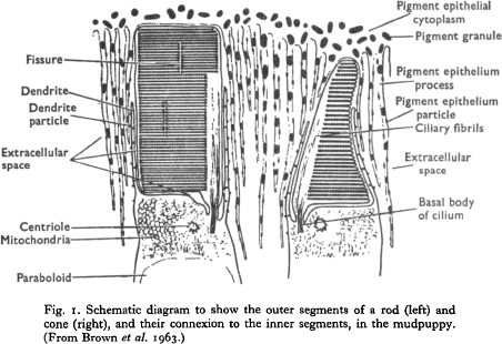
as studied by electron microscopy. Both appear to be made up of a pile of transverse paired membranes. In cones these arise by infolding of the plasma membrane, and in rods they have probably arisen in a similar way, but each pair of membranes is sealed around the edge so as to form a closed double-membrane disc (Sjöstrand, 1961). Because of the universal lamellation within the rod and cone outer segments, it looks as if there were no appreciable intracellular space, but yet Toyoda, Nosaki & Tomita (1969), and Toyoda et al. (1970) were successful in intracellular recording from the outer segments of single rods of the nocturnal gecko and frog.

Type
Research Article
Copyright
Copyright © Cambridge University Press 1970

Access options

Get access to the full version of this content by using one of the access options below. (Log in options will check for institutional or personal access. Content may require purchase if you do not have access.)

References

Arden, G. B. & Ikeda, H. (1966). Effects of hereditary degeneration of the retina on the early receptor potential and the corneo-fundal potential of the rat eye. Vision Res. 6, 171184.CrossRefGoogle ScholarPubMed
Bortoff, A. (1964). Localization of slow potential responses in the Necturus retina. Vision Res. 4, 627635.Google Scholar
Bortoff, A. & Norton, A. L. (1965 a). Simultaneous recording of photoreceptor potentials and the P III component of the ERG. Vision Res. 5, 527533.CrossRefGoogle Scholar
Bortoff, A. & Norton, A. L. (1965 b). Positive and negative potential responses associated with vertebrate photoreceptor cells. Nature, Lond. 206, 626627.CrossRefGoogle ScholarPubMed
Bortoff, A. & Norton, A. L. (1967). An electrical model of the vertebrate photoreceptor cell. Vision Res. 7, 253263.CrossRefGoogle ScholarPubMed
Brindley, G. S. & Gardner-Medwin, A. R. (1966). The origin of the early receptor potential of the retina. J. Pkysiol., Lond. 182, 185194CrossRefGoogle ScholarPubMed
Brown, K. T. (1966). The analysis of ERG and the origin of its components. Jap. J. Ophthal. 10, Suppl. (Proc. 4th ISCERG Symp.), 130140.Google Scholar
Brown, K. T. (1968). The electroretinogram: its components and their origins. Vision Res. 8, 633677.Google Scholar
Brown, K. T. & Murakami, M. (1964). A new receptor potential of the monkey retina with no detectable latency. Nature, Lond. 201, 626628.CrossRefGoogle ScholarPubMed
Brown, K. T. & Watanabe, K. (1962 a). Isolation and identification of a receptor potential from the pure cone fovea of the monkey retina. Nature, Lond. 193, 958960.Google Scholar
Brown, K. T. & Watanabe, K. (1962 b). Rod receptor potential from the retina of the night monkey. Nature, Lond. 196, 547550.CrossRefGoogle ScholarPubMed
Brown, K. T. & Wiesel, T. N. (1961 a). Analysis of the intraretinal electroretinogram in the intact cat eye. J. Physiol., Lond. 158, 229256.CrossRefGoogle ScholarPubMed
Brown, K. T. & Wiesel, T. N. (1961 b). Localization of origins of electroretinogram components by intraretinal recording in the intact cat eye. J. Physiol, Lond. 158, 257280.CrossRefGoogle ScholarPubMed
Brown, P. K., Gibbons, I. R. & Wald, G. (1963). The visual cells and visual pigment of the mudpuppy, Necturus. J. Cell Biol. 19, 70106.Google ScholarPubMed
Cone, R. A. (1967). Early receptor potential: Photoreversible charge displacement in rhodopsin. Science, N.Y. 155, 11281131.Google Scholar
Cone, R. A. & Brown, P. K. (1967). Dependence of the early receptor potential on the orientation of rhodopsin. Science, N. Y. 156, 536.Google Scholar
Cone, R. A. & Cobbs, W. H. III (1969). Rhodopsin cycle in the living eye of the rat. Nature, Land. 221, 820822.CrossRefGoogle ScholarPubMed
Dowling, J. E. & Werblin, F. S. (1969). Organization of retina of the mudpuppy, Necturus maculosus. I. Synaptic structure. J. Neurophysiol. 32, 315338.CrossRefGoogle ScholarPubMed
Eccles, J. C. (1964). The Physiology of Synapses. Berlin: Springer–Verlag.CrossRefGoogle Scholar
Frank, R. N. & Goldsmith, T. H. (1967). Effects of cardiac glycosides on electrical activity in the isolated retina of the frog. J. Gen. Physiol. 50, 15851606.CrossRefGoogle ScholarPubMed
Furukawa, T. & Hanawa, I. (1955). Effects of some common cations on electroretinogram of the toad. Jap. J. Physiol. 5, 289300.CrossRefGoogle ScholarPubMed
Granit, R. (1933). The components of the retinal action potential and their relation to the discharge in the optic nerve. J. Physiol., Lond. 77, 207240.CrossRefGoogle Scholar
Granit, R. (1947). Sensory Mechanisms of the Retina. London: Oxford University Press.Google Scholar
Granit, R. & Helme, T. (1939). Changes in retinal excitability due to polarization and some observations on the relation between the processes in retina and nerve. J. Neurophysiol. 2, 556565.CrossRefGoogle Scholar
Granit, R. & Riddell, H. A. (1934). The electrical responses of light- and dark-adapted frog's eyes to rhythmic and continuous stimuli, J. Physiol., Lond. 81, 128.CrossRefGoogle ScholarPubMed
Hagins, W. A. (1965). Electrical signs of information flow in photoreceptors. Cold Spring Harbor Symp. quant. Biol. 30, 403417.CrossRefGoogle ScholarPubMed
Hagins, W. A. & McGaughy, R. E. (1967). Molecular and thermal origins of fast photoelectric effects in the squid retina. Science, N.Y. 157, 813816.Google Scholar
Hamasaki, D. I. (1963). The effect of sodium ion concentration on the electroretinogram of the isolated retina of the frog. J. Physiol., Lond. 167, 156168.CrossRefGoogle ScholarPubMed
Hanitzsch, R. & Trifonow, J. (1968). Intraretinal abgeleitete ERG-Komponenten der isolierten Kaninchennetzhaut. Vision Res. 8, 14451455.CrossRefGoogle Scholar
Holmgren, F. (1865). Method att objectivera effecten av ljusintryck pa retina. Upsala Läkaref. förh. 1, 177191.Google Scholar
Kaneko, A. & Hashimoto, H. (1967). Recording site of the single cone response determined by an electrode marking technique. Vision Res. 7, 847851.CrossRefGoogle ScholarPubMed
Laufer, M., Svaetichin, G., Mitarai, G., Fatehchand, R., Vallecalle, E. & Villegas, J. (1961). The effect of temperature, carbon dioxide and ammonia on the neuron-glia unit. In The Visual System: Neurophysiology and Psychophysics. (Jung, R. and Kornhuber, H., eds), pp. 457463. Berlin: Springer.Google Scholar
Liebman, P. A. (1962). In situ microspectrophotometric studies on the pigments of single retinal rods. Biophys. J. 2, 161178.CrossRefGoogle ScholarPubMed
Marks, W. B. (1965). Visual pigments of single goldfish cones. J. Physiol., Lond. 178, 1432.Google Scholar
Müller-Limmroth, W. & Blümer, H. (1957). Ueber den Einfluss von Monojodessigsäure, Natriumazid und Natriumjodat auf das Ruhepotential und das Electroretinogramm des Froschauges. Z. Biol. 109, 420439.Google Scholar
Murakami, M. & Kaneko, A. (1966). Differentiation of P III subcomponents in cold-blooded vertebrate retinas. Vision Res. 6, 627636.CrossRefGoogle Scholar
Noell, W. K. (1953). Studies on the Electrophysiology and the Metabolism of the Retina. School of Aviation Med. Rep. no. 1. Randolph Field, Texas.Google Scholar
Oikawa, T., Ogawa, T. & Motokawa, K. (1959). Origin of so-called cone action potential. J. Neurophysiol. 22, 102111.Google Scholar
Pak, W. L. (1965). Some properties of the early electrical response in the vertebrate retina. Cold Spring Harb. Symp. quant. Biol. 30, 493499.CrossRefGoogle ScholarPubMed
Pak, W. L. & Cone, R. A. (1964). Isolation and identification of the initial peak of the early receptor potential. Nature, Lond. 204, 836838.CrossRefGoogle ScholarPubMed
Penn, R. D. & Hagins, W. A. (1969). Signal transmission along retinal rods and the origin of the electroretinographic a-wave. Nature, Lond. 223, 201215.CrossRefGoogle ScholarPubMed
Purple, R. E. & Dodge, F. A. (1965). Interaction of excitation and inhibition in the eccentric cell in the eye of Limulus. Cold Spring Harb. Symp. quant. Biol. 30, 529537.Google Scholar
Rushton, W. A. H. (1959). A theoretical treatment of Fuortes's observations upon eccentric cell activity in Limulus. J. Physiol., Lond. 148, 2938.CrossRefGoogle ScholarPubMed
Schultze, M. (1866). Zur Anatomie und Physiologie der Retina. Arch. mikr. Anal. 2, 175286.CrossRefGoogle Scholar
Sillman, A. J., Ito, H. & Tomita, T. (1969 a). Studies on the mass receptor potential of the isolated frog retina. I. General properties of the response. Vision Res. 9, 14351442.CrossRefGoogle ScholarPubMed
Sillman, A. J., Ito, H. & Tomita, T. (1969 b). Studies on the mass receptor potential of the isolated frog retina. II, On the basis of the ionic mechanism. Vision Res. 9, 14431451.CrossRefGoogle ScholarPubMed
Sjöstrand, F. S. (1961). Electron microscopy of the retina. In The Structure of the Eye. (Smelser, G. K., ed.), pp. 128. New York: Academic Press.Google Scholar
Skou, J. C. (1965). Enzymic basis for active transport of Na+ and K+ across cell membrane. Physiol. Rev. 45, 596617.Google Scholar
Svaetichin, G. (1953). The cone action potential. Acta Physiol. Scand. 29, Suppl. 106, 565600.Google Scholar
Svaetichin, G., Negishi, K. & Fatehchand, R. (1965). Cellular mechanisms of a Young-Hering visual system. In Colour Vision: Ciba Foundation Symposium. (de Reuck, A. V. S. and Knight, J., eds.), pp. 178207. Boston: Little, Brown and Company.Google Scholar
Tansley, K. (1964). The gecko retina. Vision Res. 4, 3337.CrossRefGoogle ScholarPubMed
Therman, P. O. (1938). The neurophysiology of the retina in the light of chemical methods of modifying its excitability. Acta Soc. Sci. Fenn. N.S.B. II, No. 1., Helsingfors.Google Scholar
Tomita, T. (1962). A compensation circuit for coaxial and double-barreled microelectrodes. IRE Trans. Biotned. Electron. 9, 138141.CrossRefGoogle Scholar
Tomita, T. (1963). Electrical activity in the vertebrate retina. J. Opt. Soc. Am. 53, 4957.CrossRefGoogle ScholarPubMed
Tomita, T. (1964). Mechanisms subserving color coding in the vertebrate retina. Abstr. 11, C III, 1, IOPAB Int. Biophys. Meeting,Paris-Orsay. Abstr. 11, C III, 1,Google Scholar
Tomita, T. (1965). Electrophysiological study of the mechanisms subserving color coding in the fish retina. Cold Spring Harb. Symp. quant. Biol. 30, 559566.CrossRefGoogle ScholarPubMed
Tomita, T. & Kaneko, A. (1965). An intracellular coaxial microelectrode. Its construction and application. Med. Electron. Biol. Engng 3, 367376.CrossRefGoogle ScholarPubMed
Tomita, T., Kaneko, A., Murakami, M. & Pautler, E. L. (1967). Spectral response curves of single cones in the carp. Vision Res. 7, 519531.CrossRefGoogle ScholarPubMed
Tomita, T., Murakami, M., Hashimoti, Y. & Sasaki, Y. (1961). Electrical activity of single neurons in the frog's retina. In The Visual System: Neurophysiology and Psychophysics. (Jung, R. and Kornhuber, H., eds.), pp. 2431. Berlin: Springer.Google Scholar
Tomita, T. & Torihama, Y. (1956). Further study on the intraretinal action potentials and on the site of ERG generation. Jap. J. Physiol. 6, 118136.Google Scholar
Toyoda, J., Nosaki, H. & Tomita, T. (1969). Light-induced resistance changes in single photoreceptors of Necturus and Gekko. Vision Res. 9, 453463.CrossRefGoogle ScholarPubMed
Toyoda, J., Hashimoto, H., Anno, H. & Tomita, T. (1970). The rod response in the frog as studied by intracellular recording. (In preparation.)CrossRefGoogle Scholar
Trifonow, Ju. A. (1968). Study of synaptic transmission between photoreceptors and horizontal cells by means of electric stimulation of the retina (in Russian). Biophysica, Moscow 13, N 5.Google Scholar
Walls, G. L. (1934). The reptilian retina. Am. J. Ophthal. 17, 892915.Google Scholar
Werblin, F. S. (1968). Functional organization of the vertebrate retina studied by intracellular recording from the retina of the mudpuppy, Necturus maculosus. Doctoral dissertation: The Johns Hopkins University, Baltimore.Google Scholar
Werblin, F. S. & Dowling, J. E. (1969). Organization of the retina of the mudpuppy, Necturus maculosus. II. Intracellular recording. J. Neurophysiol. 32, 339355.CrossRefGoogle ScholarPubMed
Yonemura, D. & Hatta, M. (1966). Localization of the minor components of the frog's electroretinogram. Jap. J. Ophthal. 10, Suppl. (Proc. 4th ISCERG Symp.), 149154.Google Scholar