


In recent years, the need to find new bioactive molecules has raised scientific interest for the marine and freshwater environments where there are a massive variety of interesting products useful for the pharmaceutical industry. In this regard, microalgae are rich sources of PUFA, such as linoleic acid, α-linolenic acid (ALA), EPA and DHA( Reference Martins, Custodio and Barreira 1 ). These valuable fatty acids from microalgae are currently being investigated to be used as nutraceuticals and therapeutic agents. Oxylipins are a large and structurally diverse family of lipid metabolites produced by the oxidative transformation of PUFA( Reference Mosblech, Feussner and Heilmann 2 ). Oxylipins are widely distributed in nature, occurring in animals, plants, mosses, algae, bacteria and fungi, and are involved in regulating cellular development and stress responses( Reference Sasso, Pohnert and Lohr 3 ). However, only a few studies have found oxylipins derived from PUFA in green microalgae( Reference Lang and Feussner 4 ). A recent investigation of lyophilised biomass samples of the microalga Chlamydomonas debaryana has led to the isolation of a series of oxylipins, including (9Z,11E,13S,15Z)-13-hydroxyoctadeca-9,11,15-trienoic acid ((13S)-HOTE) and (9Z,11E,13S)-13-hydroxyoctadeca-9,11-dienoic acid, produced by the oxidation of ALA and linoleic acid, respectively. In the same report, these oxylipins have been shown to inhibit the production of the pro-inflammatory cytokine TNF-α in lipopolysaccharide-stimulated THP-1 monocyte-derived macrophages( Reference de Los Reyes, Ávila-Román and Ortega 5 ).
In mammals, it has been shown that oxylipins are involved in the initiation and resolution of the inflammatory response. During the acute phase, arachidonic acid is converted to pro-inflammatory eicosanoids, such as leukotriene B4 and PGE2, that recruit neutrophils to the site of injury and elicit an inflammatory response. By contrast, during the resolution of the inflammatory response, a number of anti-inflammatory and pro-resolving lipid mediators are released to promote tissue healing and clearance of micro-organisms and apoptotic cells( Reference Serhan and Petasis 6 ). These oxylipins are derived from essential n-6 and n-3 PUFA and include arachidonic acid-derived lipoxins, aspirin-triggered lipoxins, EPA-derived resolvins of the E-series, DHA-derived resolvins of the D-series, protectins and maresins( Reference Serhan and Chiang 7 ).
Although inflammation is primarily a physiological and beneficial response, non-resolving inflammatory processes can lead to chronic inflammation, which is often linked to the pathogenesis and progression of several prevalent disorders, such as inflammatory bowel diseases (IBD). IBD are a group of chronic diseases that affect the gastrointestinal tract and have been mainly subdivided into ulcerative colitis and Crohn's disease (CD)( Reference Mulder, Noble and Justinich 8 ). Although the aetiology has not been completely understood, it has been shown that a combination of genetic susceptibility factors and altered immune responses driven by microbial factors in the enteric environment are involved in the pathophysiology of these diseases( Reference Kaser, Zeissig and Blumberg 9 ). Numerous animal models of IBD have been established, which can help to obtain new insights into the pathogenesis of these diseases and may be used to test innovative approaches for therapy( Reference Neurath 10 ). Colitis induced by an intrarectal injection of 2,4,6-trinitrobenzenesulfonic acid (TNBS) is a model that resembles many of the clinical, histopathological and immune characteristics of CD in humans( Reference Morris, Beck and Herridge 11 ). It has been shown that the activation of the intestinal immune system results in the production of pro-inflammatory cytokines, such as TNF-α, which amplify the inflammatory response by activating a cascade of immune cells, such as neutrophils and macrophages. Infiltration of neutrophils results in the production of cytotoxic reactive oxygen and nitrogen species, and lytic enzymes, which cause the destruction of intestinal cell macromolecules, ultimately leading to mucosal disruption( Reference Mouzaoui, Rahim and Djerdjouri 12 ).
There is compelling evidence on the anti-inflammatory properties of exogenously administered n-3 PUFA, such EPA, DHA and ALA, in experimental models of IBD( Reference Marion-Letellier, Savoye and Beck 13 ). These beneficial effects have been associated with a reduction in the production of eicosanoids such as PGE2 ( Reference Nieto, Fernandez and Torres 14 ) and leukotriene B4, and the down-regulation of pro-inflammatory cytokines, such as TNF-α( Reference Camuesco, Galvez and Nieto 15 ) and IL-1β( Reference Cho, Chi and Chun 16 ), and adhesion molecules, including intercellular adhesion molecule 1, vascular cell adhesion molecule 1 and vascular endothelial growth factor receptor 2( Reference Ibrahim, Aziz and Hassan 17 ). It has also been reported that PUFA suppress inducible enzymes such as cyclo-oxygenase 2 (COX-2) and inducible NO synthase (iNOS)( Reference Camuesco, Galvez and Nieto 15 , Reference Hassan, Ibrahim and Mbodji 18 ), as well as reduce oxidative stress in experimental colitis( Reference Hassan, Ibrahim and Mbodji 18 ). Furthermore, n-3 PUFA-derived oxylipins, such as resolvin E1 (RvE1), D-series resolvins and maresin 1, have been shown to exert important positive effects on dextran sulphate sodium (DSS)- and TNBS-induced colitis through the inhibition of neutrophil infiltration and the production of inflammatory mediators in colonic tissue( Reference Arita, Yoshida and Hong 19 ).
To further explore the beneficial effects of PUFA-derived oxylipins on IBD, we investigated the effects of the oxylipin-containing lyophilised biomass of the microalga C. debaryana and its major oxylipin constituent, (13S)-HOTE, on an experimental model of colitis in rats. In the present study, we report that these products can effectively ameliorate TNBS-induced colitis by increasing mucus production, inhibiting neutrophil infiltration and TNF-α release, as well as suppressing COX-2 and iNOS expression in the colonic tissue of rats.
Experimental methods
Experimental animals
A total of sixty-eight male Wistar rats supplied by Animal Services of the University of Seville, Spain, weighing 200–250 g, aged 5–6 weeks, were included in the present study. The rats were housed in type IV cages (n 4–5 rats per cage), containing wood shavings and wire-net floors. They were maintained on a regular 12 h light–12 h dark cycle in a temperature (24–25°C)- and humidity (70–75 %)-controlled room, and were allowed free access to tap water and a normal laboratory diet (Panlab, Barcelona, Spain). The rats were allowed to acclimatise for 1 week before the commencement of the experiments. They were deprived of food for 12 h before the induction of colitis, but were allowed free access to tap water throughout the study. They were randomly assigned to groups of eight to ten animals in a blinded fashion. The experiments followed a protocol approved by the Animal Ethics Committee of the University of Seville (P09-AGR-5185), and all experiments were in accordance with the recommendations of the European Union regarding animal experimentation (Directive of the European Council 2010/63/EU).
Induction of colitis and treatments
Colitis was induced according to the procedure described by Morris et al. ( Reference Morris, Beck and Herridge 11 ). Briefly, the rats were slightly anaesthetised with 12 % chloral hydrate by the intraperitoneal route following a 12 h fast, and then for enteral feeding, a medical-grade polyurethane cannula (external diameter 2 mm) was inserted into the anus and the tip was advanced to 8 cm proximal to the anus verge. To induce acute colitis, TNBS (Sigma Chemical Company) dissolved in ethanol (50 %, v/v) was instilled into the colon through the cannula (10 mg in a total volume of 0·25 ml). A reference control group was included for comparison with the TNBS-induced group; the sham group received physiological saline instead of the TNBS solution in a comparable volume. Following the instillation of the hapten, the rats were maintained in a head-down position for a few minutes (1–2 min) to prevent leakage of the intracolonic instillate.
The lyophilised biomass sample of the microalga C. debaryana (ITC09-1702-12) was provided by Instituto Tecnológico de Canarias (Spain). The oxylipin (13S)-HOTE was isolated from the microalgal biomass as described previously( Reference de Los Reyes, Ávila-Román and Ortega 5 ). Briefly, a sample of the lyophilised biomass was extracted with acetone–methanol (1:1), and the resulting extract was subjected to column chromatography on silica gel. Fractions eluted with hexane–diethyl ether (3:7), diethyl ether and chloroform–methanol (9:1, 8:2) were separated over reversed-phase cartridges (solid phase extraction RP-18) and then by normal-phase HPLC to obtain pure oxylipins.
The lyophilised microalgal biomass was emulsified in 0·9 % saline solution and < 5 % Tween-80 (Sigma Chemical Company), and administered by the oral route (300 and 600 mg/kg). These doses were chosen according to preliminary experiments with a reduced number of animals (J Ávila-Román and E Talero, unpublished results). The two doses of the lyophilised microalga assayed (300 and 600 mg/kg) contained 0·05 and 0·1 mg of (13S)-HOTE, respectively. The isolated compound (13S)-HOTE was suspended in 0·9 % saline solution and < 5 % Tween-80, and administered by the oral route (0·1, 0·5 and 1 mg/kg). Both the lyophilised biomass and (13S)-HOTE were administered in a volume of 1 ml/100 g body weight 48, 24 and 1 h before, and 24 h after the induction of colitis. The sham and TNBS groups also received the vehicle by the oral route. The body weight and stool consistency of rats were examined. The rats were then killed using an overdose of the anaesthetic 48 h after the induction of colitis.
Assessment of colitis
The severity of colitis was evaluated by an independent observer who was blinded to the treatment. For each animal, the distal 10 cm portion of the colon was removed and cut longitudinally, slightly cleaned in physiological saline to remove faecal residues and weighed. Macroscopic damage was quantified by measuring the extent of the lesions in the distal colon (cm2). The presence of adhesions (score 0–2) and/or stool consistency (score 0–1) were evaluated according to the criteria of Bobin-Dubigeon et al. ( Reference Bobin-Dubigeon, Collin and Grimaud 20 ) with slight modifications. Photographs taken from colon samples were digitised using a Kodak D290 Zoom camera (Eastman Kodak Company). Pieces of the colon were collected and frozen in liquid N2 for the measurement of biochemical parameters.
Histological studies
Tissue samples from the distal colon of each animal were fixed in 4 % buffered paraformaldehyde, dehydrated by increasing concentrations of ethanol and embedded in paraffin. Thereafter, sections of tissue were cut at 5 mm on a rotary microtome (Leica Microsystems), mounted on clean glass slides and dried overnight at 37°C. The sections were cleared, hydrated, and stained with haematoxylin and eosin, and alcian blue for histological evaluation of colonic damage and mucus content, respectively, according to standard protocols, and the slides were coded to prevent observer bias during evaluation. All tissue sections were examined under an Olympus BH-2 microscope (GMI Inc.) for characterisation of histopathological changes. Mucin density was quantified using Scientific Imaging Systems (Biophotonics ImageJ Analysis Software; National Institutes of Health) to study the preservation, and mucus production in colonic mucosa.
Assessment of leucocyte involvement
Myeloperoxidase (MPO) activity was assessed as an index of neutrophil infiltration according to the method of Grisham et al. ( Reference Grisham, Benoit and Granger 21 ). Samples were excised from each animal, and rapidly rinsed with ice-cold saline, blotted dry and frozen at − 80°C. The tissue was thawed, weighed and homogenised in ten volumes of 50 mm-PBS (pH 7·4). The homogenate was centrifuged at 20 000 g for 20 min at 4°C. The pellet was again homogenised in ten volumes of 50 mm-PBS (pH 6·0) containing hexadecyl trimethylammonium bromide (0·5 %) and 10 mm-EDTA. This homogenate was subjected to one cycle of freezing/thawing and a brief period of sonication. A sample of the homogenate (50 μl) was added to a ninety-six-well microplate and incubated at 37°C for 3 min with a mixture containing o-dianisidine dihydrochloride (0·067 %), hexadecyl trimethylammonium bromide (0·5 %) and 0·3 mm-H2O2. The changes in absorbance at 655 nm were measured with a microplate reader (Labysistem Multiskan EX, Thermo Scientific). One unit of MPO activity was defined as the amount of enzyme present that produced a change in absorbance of 1·0 unit/min at 37°C in the final reaction volume containing the acetate. Results are expressed as units/mg tissue.
Measurement of TNF-α
Distal colon samples were weighed and homogenised at 4°C, after thawing, in 0·3 ml PBS (pH 7·2) and 1 % bovine serum albumin containing 0·01 mg/ml of leupeptin, 0·01 mg/ml of pepstatin, 0·01 mg/ml of aprotinin and 1 mm-phenylmethylsulfonyl fluoride. Then, the homogenates were centrifuged at 12 000 g for 10 min. TNF-α levels in colonic tissues were measured by a quantitative enzyme immunoassay kit (Diaclone), according to the manufacturer's protocol. TNF-α values are expressed as ng/mg tissue.
Isolation of cytoplasmic proteins and Western blot assay
Frozen colonic tissues were weighed and homogenised in ice-cold buffer (50 mm-Tris–HCl, pH 7·5, 8 mm-MgCl2, 5 mm-ethylene glycol bis(2-aminoethyl ether)-N, N, N′N′-tetraacetic acid, 0·5 mm-EDTA, 0·01 mg/ml of leupeptin, 0·01 mg/ml of pepstatin, 0·01 mg/ml of aprotinin, 1 mm-phenylmethylsulfonyl fluoride and 250 mm-NaCl). The homogenates were centrifuged at 12 000 g for 15 min at 4°C, and the supernatants were collected and stored at − 80°C. Protein concentration of the homogenates was determined following the colorimetric method of Bradford( Reference Bradford 22 ). Aliquots of the supernatants containing equal amounts of proteins (50 μg) were separated on 10 % acrylamide gel by SDS–PAGE. Subsequently, the proteins were electrophoretically transferred onto a nitrocellulose membrane and incubated with specific primary antibodies: rabbit anti-COX-2 and anti-iNOS (Cayman Chemical) at a dilution of 1:1000. Each membrane was washed three times for 15 min and incubated with the secondary horseradish peroxidase-linked anti-rabbit (Pierce Chemical). To prove equal loading, the blots were analysed for β-actin expression using an anti-β-actin antibody (Sigma-Aldrich). Immunodetection was performed using an enhanced chemiluminescence light-detecting kit (Super-Signal West Pico Chemiluminescent Substrate; Pierce Chemical). Densitometric data were studied following normalisation to the control (housekeeping gene). The signals were analysed and quantified with a Scientific Imaging Systems (Biophotonics ImageJ Analysis Software; National Institutes of Health).
Statistical analyses
Data are expressed as means with their standard errors. Statistical analyses were performed using the IBM SPSS Statistics 22.0 software. In all cases, the Shapiro–Wilk test was used to verify the normality of the data. Statistical significance between the two control groups (sham v. TNBS) was determined by Student's t test. The Mann–Whitney U test was chosen for non-parametric values. Statistical differences between several groups were determined by one-way ANOVA followed by Bonferroni's post hoc test for parametric data. When variances were heterogeneous, Welch's test followed by Tamhane's test was conducted. Non-parametric data were analysed by the Kruskal–Wallis test for multiple comparisons. P values < 0·05 were considered statistically significant. In the histological experiment, results shown are representative of at least three experiments performed on different days. The statistical test used for individual analyses is provided in the figure legends.
Results
Lyophilised microalga and (9Z,11E,13S,15Z)-13-hydroxyoctadeca-9,11,15-trienoic acid protect rats against 2,4,6-trinitrobenzenesulfonic acid-induced colitis
The data from rats with colitis induced by 10 mg/animal of TNBS following an acute study are shown in Figs. 1 and 2 and in Table 1. The administration of TNBS by rectal instillation in rats resulted in severe colitis. As shown in Table 1, the presence of diarrhoea (P< 0·001) accompanied by a marked body-weight loss (P< 0·001) was evident in TNBS-induced rats. This experimental group also experienced a significant increase in colonic weight:length ratio, a marker of tissue inflammation (P< 0·001), and greater adhesions to adjacent organs than the sham group (P< 0·001). Macroscopic inspection of the colon and rectum provided evidence for severe colonic mucosal damage, with deep haemorrhagic ulcerations and necrosis (Fig. 1(b)) in comparison with the sham group (Fig. 1(a)). Macroscopic lesions in the distal colon were quantified and the affected area was 8·6 (sem 0·5) cm2 (P< 0·001 v. sham group) (Fig. 1(h)).

Fig. 1 Lyophilised microalgal biomass and (9Z,11E,13S,15Z)-13-hydroxyoctadeca-9,11,15-trienoic acid ((13S)-HOTE) administration attenuate trinitrobenzenesulfonic acid (TNBS)-induced acute colitis. (a–g) Representative macroscopic appearance of the colon of rats treated with the lyophilised microalga ((c) L-300 and (d) L-600 mg/kg by the oral route) and (13S)-HOTE ((e) H-0·1, (f) H-0·5 and (g) H-1 mg/kg by the oral route), compared with the (a) sham group and (b) TNBS group. (h) Colon macroscopic damage was measured as indicated in the ‘Experimental methods’ section. Values are means, with their standard errors represented by vertical bars. Mean value was significantly different from that of the TNBS group: * P< 0·05; ** P< 0·01; *** P< 0·001 (one-way ANOVA followed by Bonferroni's post hoc test). ††† Mean value was significantly different from that of the sham group (P< 0·001; Student's t test). L-300, 300 mg lyophilised microalga Chlamydomonas debaryana/kg animal; L600, 600 mg lyophilised microalga C. debaryana/kg animal; H-0·1, 0·1 mg (13S)-HOTE/kg animal; H-0·5, 0·5 mg (13S)-HOTE/kg animal; H-1, 1 mg (13S)-HOTE/kg animal.

Fig. 2 Lyophilised microalgal biomass and (9Z,11E,13S,15Z)-13-hydroxyoctadeca-9,11,15-trienoic acid ((13S)-HOTE) administration attenuate microscopic colon damage induced by trinitrobenzenesulfonic acid (TNBS, 10 mg/animal) and improve mucus accumulation inside the goblet cells. Histological appearance of rat colonic mucosa after haematoxylin and eosin staining and alcian blue staining: (a, b) sham group; (c, d) TNBS group; (e, f) lyophilised microalgal biomass (600 mg/kg by the oral route) and (g, h) (13S)-HOTE (1 mg/kg by the oral route). Original magnification 200 × . (i) Mucin density was studied following normalisation to the sham group. Results are representative of three experiments performed on different samples. Values are means, with their standard errors represented by vertical bars. ** Mean value was significantly different from that of the TNBS group (P< 0·01; one-way ANOVA followed by Bonferroni's post hoc test). ††† Mean value was significantly different from that of the sham group (P< 0·001; Student's t test). L-300, 300 mg lyophilised microalga Chlamydomonas debaryana/kg animal; L600, 600 mg lyophilised microalga C. debaryana/kg animal; H-0·1, 0·1 mg (13S)-HOTE/kg animal; H-0·5, 0·5 mg (13S)-HOTE/kg animal; H-1, 1 mg (13S)-HOTE/kg animal.
Table 1 Effect of the lyophilised microalgal biomass and (9Z,11E,13S,15Z)-13-hydroxyoctadeca-9,11,15-trienoic acid ((13S)-HOTE) administration on the clinical signs of colitis induced by trinitrobenzenesulfonic acid (TNBS; 10 mg/animal)‡ (Mean values with their standard errors)
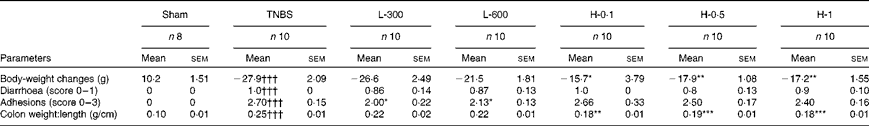
L-300, 300 mg lyophilised microalga Chlamydomonas debaryana/kg animal; L-600, 600 mg lyophilised microalga C. debaryana/kg animal; H-0·1, 0·1 mg (13S)-HOTE/kg animal; H-0·5, 0·5 mg (13S)-HOTE/kg animal; H-1, 1 mg (13S)-HOTE/kg animal.
Mean value was significantly different from that of the TNBS group: * P< 0·05, ** P< 0·01, *** P< 0·001. ANOVA P value, followed by Bonferroni's post hoc test, comparing the TNBS and lyophilised groups are shown. For body weight changes, P values for Welch's test followed by Tamhane's test comparing the TNBS and (13S)-HOTE groups are shown. For colon weight:length, P values for the Kruskal–Wallis test comparing the TNBS and (13S)-HOTE-treated rats are shown.
††† Mean value was significantly different from that of the sham group (P< 0·001; Student's t test).
‡ Quantified parameters in rats with colitis induced by TNBS and treated with a lyophilised biomass sample of the microalga C. debaryana (L-300 and L-600 mg/kg animal) and the oxylipin (13S)-HOTE (H-0·1, H-0·5 and H-1 mg/kg animal) following an acute approach.
Administration of (13S)-HOTE treatment at the three doses used (0·1, 0·5 and 1 mg/kg) significantly attenuated the loss in body weight and the weight:length ratio of the rat colon (Table 1). The presence of adhesions to adjacent organs was only reduced in lyophilisate-treated rats. With respect to diarrhoea, no significant changes were observed with any of the treatments assayed. In addition, the extent and severity of colonic injury was significantly reduced in rats treated with 600 mg/kg of lyophilised microalga (Fig. 1(d)) and 0·1, 0·5 and 1 mg/kg of (13S)-HOTE (Figs. 1(e)–(g)). In fact, (13S)-HOTE was able to reduce macroscopic damage down to 5·2 (sem 0·6) cm2 with the highest dose (P< 0·001 v. TNBS group) (Fig. 1(h)).
Lyophilised microalga and (9Z,11E,13S,15Z)-13-hydroxyoctadeca-9,11,15-trienoic acid alleviate microscopic colon damage and increase mucus production
The histological analysis of the colon of sham rats revealed typical features of a normal structure (Fig. 2(a)). Consistent with macroscopic changes, TNBS-induced rats exhibited marked transmural inflammation involving all layers of the bowel wall. Extensive granulation tissue with the presence of massive neutrophilic infiltration, fibroblasts and lymphocytes was also apparent, mainly in the mucosa and submucosa. Necrosis of the epithelium, distortion of crypts and partial destruction of the glands were also detected in these animals (Fig. 2(c)). Alcian blue staining, which detects acid mucin-positive goblet cells, revealed remarkable mucin depletion in the ulcerative areas of TNBS animals (Fig. 2(d)) compared with sham rats (Fig. 2(b)). In contrast, the histological sections of the lyophilised biomass (Fig. 2(e)) and (13S)-HOTE-treated animals (Fig. 2(g)) showed an improvement in the microscopic features of colitis with all the doses used, evidenced by a preservation of the colonic mucosal structure and a reduction of inflammatory cells in the lamina propria when compared with the TNBS group. Furthermore, alcian blue-positive goblet cells were clearly observed in the preserved regions of the mucosal layer in rats treated with lyophilised biomass (Fig. 2(f)) or (13S)-HOTE (Fig. 2(h)). These observations were confirmed by the quantification of mucin density (Fig. 2(i)). The lyophilised biomass (600 mg lyophilised microalga C. debaryana/kg animal) and (13S-HOTE) showed higher mucus production in comparison with the TNBS group (P <0·01).
Lyophilised microalga and (9Z,11E,13S,15Z)-13-hydroxyoctadeca-9,11,15-trienoic acid decrease neutrophil infiltration and colonic TNF-α production
Neutrophil infiltration detected in the histological examination of the colon from TNBS-induced rats correlated with increased colonic MPO activity, an established marker for inflammatory cell infiltration into the colon (1·28 (sem 0·12) units/mg tissue, P< 0·01 v. sham group) (Fig. 3(a)). Treatment with lyophilised microalgal biomass (300 and 600 mg/kg), or (13S)-HOTE (0·5 and 1 mg/kg) significantly reduced the degree of polymorphonuclear neutrophil infiltration in colonic tissue (lyophilised microalgae: 0·51 (sem 0·10) and 0·65 (sem 0·06) units/mg tissue, P< 0·01 v. TNBS group; (13S)-HOTE: 0·50 (sem 0·23) and 0·22 (sem 0·04) units/mg tissue, P< 0·05 and P< 0·01, respectively v. TNBS group) (Fig. 3(a)). Colonic injury by TNBS administration was also characterised by a dramatic rise in TNF-α concentration to 2·52 (sem 0·43) ng/mg tissue compared with normal rats (0·92 (sem 0·13) ng/mg tissue, P< 0·01). In contrast, the levels of this cytokine were significantly lower in rats treated with either the lyophilised microalga or (13S)-HOTE at all the doses assayed (Fig. 3(b)).

Fig. 3 Lyophilised microalgal biomass and (9Z,11E,13S,15Z)-13-hydroxyoctadeca-9,11,15-trienoic acid ((13S)-HOTE) acute administration reduce the infiltration of leucocytes and the production of the pro-inflammatory cytokine TNF-α in trinitrobenzenesulfonic acid (TNBS)-induced colitis. (a) Myeloperoxidase (MPO) activity and (b) TNF-α levels were quantified in rats treated with TNBS alone or rats receiving TNBS plus lyophilised microalgal biomass (L-300 and L-600 mg/kg by the oral route) or (13S)-HOTE (H-0·1, H-0·5 and H-1 mg/kg by the oral route). The sham group received physiological saline instead of the TNBS solution in an equal volume. Values are means, with their standard errors represented by vertical bars. (a) Mean value was significantly different from that of the TNBS group: * P< 0·05; ** P< 0·01 (one-way ANOVA followed by Bonferroni's post hoc test for lyophilised-treated groups v. TNBS group; Kruskal–Wallis test for (13S)-HOTE-treated groups v. TNBS group). †† Mean value was significantly different from that of the sham group (P< 0·01; Mann–Whitney U test). (b) ** Mean value was significantly different from that of the TNBS group (P< 0·01; one-way ANOVA followed by Bonferroni's post hoc test for lyophilised-treated groups v. TNBS group; Welch's test followed by Tamhane's test for (13S)-HOTE-treated groups v. TNBS group). †† Mean value was significantly different from that of the sham group (P< 0·01; Student's t test). L-300, 300 mg lyophilised microalga Chlamydomonas debaryana/kg animal; L600, 600 mg lyophilised microalga C. debaryana/kg animal; H-0·1, 0·1 mg (13S)-HOTE/kg animal; H-0·5, 0·5 mg (13S)-HOTE/kg animal; H-1, 1 mg (13S)-HOTE/kg animal.
Lyophilised microalga and (9Z,11E,13S,15Z)-13-hydroxyoctadeca-9,11,15-trienoic acid down-regulate the expression of cyclo-oxygenase 2 and inducible nitric oxide synthase in colonic mucosa
We examined COX-2 and iNOS expression by Western blotting of cytosolic extracts from colonic mucosa (Fig. 4(a)). Exposure of the colon to TNBS induced a pronounced increase in COX-2 and iNOS protein levels compared with sham animals (P< 0·01 and P< 0·001, respectively). The treatment with the lyophilised microalga or (13S)-HOTE resulted in a significant decrease in the levels of these enzymes. Interestingly, iNOS expression drastically decreased under basal levels with the highest dose of (13S)-HOTE (Fig. 4(b)).

Fig. 4 Lyophilised microalgal biomass and (9Z,11E,13S,15Z)-13-hydroxyoctadeca-9,11,15-trienoic acid ((13S)-HOTE) acute administration reduce colonic protein levels of cyclo-oxygenase 2 (COX-2, □) and inducible nitric oxide synthase (iNOS, ■) enzymes in trinitrobenzenesulfonic acid (TNBS)-induced colitis. (a) Representative Western blot analysis of COX-2 and iNOS proteins. (b) Densitometric data were studied following normalisation to the control (housekeeping gene, β-actin). Results are representative of three experiments performed on different samples. Values are means, with their standard errors represented by vertical bars. Mean value was significantly different from that of the TNBS group: * P< 0·05; ** P< 0·01 (one-way ANOVA followed by Bonferroni's post hoc test). Mean value was significantly different from that of the sham group: †† P< 0·01; ††† P< 0·001 (Student's t test). L-300, 300 mg lyophilised microalga Chlamydomonas debaryana/kg animal; L600, 600 mg lyophilised microalga C. debaryana/kg animal; H-0·1, 0·1 mg (13S)-HOTE/kg animal; H-0·5, 0·5 mg (13S)-HOTE/kg animal; H-1, 1 mg (13S)-HOTE/kg animal.
Discussion and conclusions
Ulcerative colitis and CD are major forms of IBD, a disease that affects millions of people worldwide and is characterised by chronic uncontrolled inflammation of intestinal mucosa. IBD therapies have modest results for long-term management and significant side effects. It has been reported that diet and nutritional factors play a key role in IBD, and, thus, developing nutritional interventions against this pathology remains important( Reference Hou, Lee and Lewis 23 ). In this regard, n-3 PUFA have been suggested as a treatment for IBD, because of their anti-inflammatory effects( Reference Marion-Letellier, Savoye and Beck 13 ). PUFA are precursors of oxylipins, lipid mediators that are extremely important for the resolution of many inflammatory disorders( Reference Serhan and Chiang 7 ). Microalgae have been proposed as an interesting natural source in the search for novel bioactive compounds and functional food products( Reference Plaza, Herrero and Cifuentes 24 ). It has been shown that microalgal species, being a promising source of n-3 PUFA and derived oxylipins, accumulate high amounts of lipids( Reference Martins, Custodio and Barreira 1 ). In this context, the aim of the present study was to evaluate the possible anti-inflammatory effects of the oxylipin-containing lyophilised biomass of the microalga C. debaryana and the ALA-derived oxylipin (13S)-HOTE in an experimental model of colitis. We have shown for the first time that lyophilised microalgal biomass or (13S)-HOTE exerts protective effects against TNBS-induced colitis.
The present results clearly demonstrated that the intracolonic administration of 10 mg TNBS caused an acute colitis in rats, characterised by the presence of diarrhoea, colonic shortening and an increase in colonic wall thickness, produced by a more extensive inflammatory cell infiltrate in the mucosa and submucosa. Treatment of rats with lyophilised microalgal biomass or (13S)-HOTE inhibited body-weight loss, colon shortening and the extent of intestinal inflammation. Microscopic analysis of the colon confirmed the data from the macroscopic study reflecting the attenuation of mucosal disruption and oedema after the treatment with the lyophilised microalga and (13S)-HOTE. These findings are in agreement with previous studies which showed that PUFA are effective in controlling disease activity in experimental colitis. In this line, dietary olive oil enriched with EPA and DHA, or oral administration of pure DHA, resulted in a significant inhibition of the incidence of diarrhoea, body-weight loss, colon shortening and histological damage when colitis was induced by incorporating DSS in drinking-water( Reference Camuesco, Galvez and Nieto 15 , Reference Cho, Chi and Chun 16 ). Likewise, treatment with n-3 PUFA-derived oxylipins, such as RvE1, D-series resolvins and maresin 1, greatly improved clinical signs of colitis and colonic damage induced by DSS or TNBS( Reference Arita, Yoshida and Hong 19 , Reference Ishida, Yoshida and Arita 25 – Reference Marcon, Bento and Dutra 27 ). In fact, RvE1 has been shown to promote the resolution of the inflammatory response through epithelial intestinal cells by the activation of intestinal alkaline phosphatase, the detoxification of lipopolysaccharide and the reduction of lipopolysaccharide-induced inflammation by the down-regulation of the NF-κB signalling pathway( Reference Campbell Eric, MacManus Christopher and Kominsky Douglas 28 ). These findings suggest that intestinal alkaline phosphatase is a major regulatory enzyme of gut homeostasis and health( Reference Lallès 29 ). Interestingly, it has also been reported that lipoxin A4, an arachidonic acid-derived lipoxin, as well as n-6 docosapentaenoic acid-derived resolvins, protected mice from DSS-induced colitis( Reference Gewirtz, Collier-Hyams and Young 30 , Reference Chiu, Gomolka and Dierkes 31 ).
Intestinal goblet cells produce intestinal mucus that completely fills the crypts. Mucins, the principal components of the mucus, are responsible for its high viscosity and forming a gel-like mucus layer, which serves as a barrier to protect the intestinal epithelial layer from the injurious effects of luminal stimulants( Reference Ermund, Schutte and Johansson 32 ). Altered goblet cell physiology is a hallmark of IBD pathology( Reference Boltin, Perets and Vilkin 33 ). Individuals with CD or ulcerative colitis have been shown to have decreased numbers of goblet cells and reduced mucus thickness( Reference Heazlewood, Cook and Eri 34 ). The present study confirmed mucin-depleted crypts in rats with TNBS-induced colitis, as attested by the loss of alcian blue-stained goblet cells. Interestingly, the lyophilised microalgal biomass or (13S)-HOTE treatment improved mucus accumulation inside the goblet cells, which suggests a protective effect of these compounds on colonic epithelial damage that occurs in colitis. In a similar way, fish oil diet enriched with n-3 PUFA increased the number of goblet cells in colonic mucosa from TNBS-induced rats( Reference Nieto, Torres and Rios 35 ).
TNBS-induced colitis provokes a dominant T helper cell type 1 response, immunologically similar to human CD, requiring T-cell activation as one of the initiating events that subsequently leads to macrophage/neutrophil recruitment and activation( Reference Zhang, Zhou and Zhou 36 ). Since the cellular infiltrate has pathogenic roles in IBD, its control is extremely important for the attenuation of this disease. Tissue levels of MPO activity are used as a quantitative measure of neutrophil inflammatory response in a variety of experimental studies( Reference Kolgazi, Uslu and Yuksel 37 ). Our findings showed that oral administration of lyophilised microalga or (13S)-HOTE ameliorated polymorphonuclear infiltration into the colon, as evidenced by the suppression of colonic MPO activity as well as the improvement of histological features. The immune cells participate actively in the mucosal immune and inflammatory responses by producing pro-inflammatory cytokines such as TNF-α, which play a crucial role in the initiation and maintenance of mucosal inflammation and immunity in IBD( Reference Amrouche-Mekkioui and Djerdjouri 38 ). This cytokine is involved in many cell processes including apoptotic cell death, metabolism, inflammation, thrombosis and fibrinolysis( Reference Mouzaoui, Rahim and Djerdjouri 12 ). One major action of TNF-α is the induction of adhesion molecule and chemokine expression, resulting in an influx of inflammatory cells. A previous study revealed high levels of TNF-α in the serum and colonic mucosa of patients with IBD( Reference Komatsu, Kobayashi and Saito 39 ). The anti-inflammatory effects of PUFA, mediated by inhibiting the release of TNF-α, have previously been reported in various experimental models of colitis( Reference Camuesco, Galvez and Nieto 15 , Reference Cho, Chi and Chun 16 , Reference Hassan, Ibrahim and Mbodji 18 ). Likewise, treatment with RvE1, resolvins of the D series and maresin 1 suppressed colonic mRNA expression and protein levels of TNF-α in TNBS- and DSS-induced colitis( Reference Arita, Yoshida and Hong 19 , Reference Ishida, Yoshida and Arita 25 – Reference Marcon, Bento and Dutra 27 ). Similarly, administration of lyophilised microalgal biomass or (13S)-HOTE consistently decreased the release of TNF-α in the colon. The present results suggest that these lipid products may contribute to the prevention of cell infiltration by suppressing the levels of TNF-α, and thus, in turn, ameliorate colon inflammation.
COX-2 and iNOS are immediate-early response genes normally absent from most cells, but are induced mainly at the sites of inflammation in response to inflammatory stimuli including cytokines such as TNF-α( Reference Kanwar, Kanwar and Burrow 40 , Reference Wang and Dubois 41 ). These two pro-inflammatory enzymes are up-regulated in experimental colitis( Reference Talero, Sanchez-Fidalgo and de la Lastra 42 ) and in active human IBD( Reference Cirillo, Sarnelli and Esposito 43 , Reference Fratila and Ilias 44 ), generating a large number of products implicated in oxidative damage and inflammation( Reference Yasukawa, Tokuda and Tun 45 ). In the present study, colonic damage was associated with a high expression of both COX-2 and iNOS proteins. However, lyophilised microalga or (13S)-HOTE administered orally significantly down-regulated the expression of these proteins in the colonic tissue after acute TNBS administration. Consistent with these findings, a previous paper demonstrated a reduction in colonic mRNA levels of COX-2 and iNOS in colitic mice that received RvE1( Reference Arita, Yoshida and Hong 19 ). Similar results were found by Bento et al. ( Reference Bento, Claudino and Dutra 26 ) after administration of resolvins of the D series. Interestingly, these authors have shown that the anti-inflammatory effects of these PUFA-derived mediators were associated with the inhibition of NF-κB, which has been shown to be activated in experimental colitis.
In conclusion, we have shown for the first time the preventive effects of a lyophilised biomass sample of the microalga C. debaryana and the ALA-derived oxylipin (13S)-HOTE, in a TNBS-induced colitis model in rats. These actions may be associated with an enhanced colonic mucus release, a polymorphonuclear reduction in PMN infiltration, and TNF-α levels, as well as the suppression of COX-2 and iNOS expression in colonic tissue. Future studies are needed to expand the vision of the effects and mechanisms by which these lipid products improve experimental colitis. These investigations will support the potential use of this microalga, or derived oxylipins, as nutraceuticals in the treatment of the active phase of IBD.
Acknowledgements
The authors thank A. de la Jara (Instituto Tecnológico de Canarias, Spain) for providing the lyophilised microalgal samples, A. Fernández-Palacín for the statistical data analysis, and Centro de Investigación, Tecnología e Innovación of the University of Seville (CITIUS) for providing technical assistance.
The present study was supported by Secretaría de Innovación, Ministerio de Ciencia Innovación, Gobierno de España (grant no. IPT-2011-1370-060000)
The authors’ contributions are as follows: E. T., E. Z., S. G.-M. and V. M. designed the study protocol; C. D. L. R. and E. Z. extracted the lyophilised microalgal biomass and obtained the oxylipin (13S)-HOTE; J. A.-R., E. T. and A. A. conducted the in vivo experiments; J. A.-R. and E. T. performed the histological experiments and analysed the data; J. A.-R. and E. T. wrote the draft of the manuscript. All authors critically reviewed and approved the final version of the manuscript.
None of the authors has any conflict of interest to declare.


