Hostname: page-component-8448b6f56d-c4f8m Total loading time: 0 Render date: 2024-04-24T22:00:55.477Z Has data issue: false hasContentIssue false

Quantifying plant cell-wall failure in vivo using nanoindentation

Published online by Cambridge University Press:  28 August 2014

Elham Forouzesh
Affiliation:
Mechanical and Materials Engineering, University of Nebraska-Lincoln, W342 Nebraska Hall, Lincoln, Nebraska 68588-0526
Ashwani K. Goel
Affiliation:
Mechanical and Materials Engineering, University of Nebraska-Lincoln, W342 Nebraska Hall, Lincoln, Nebraska 68588-0526
Joseph A. Turner*
Affiliation:
Mechanical and Materials Engineering, University of Nebraska-Lincoln, W342 Nebraska Hall, Lincoln, Nebraska 68588-0526
*
Address all correspondence to Joseph A. Turner atjaturner@unl.edu
Get access

Abstract

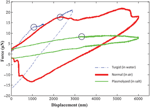
Nanoindentation experiments have been carried on Arabidopsis thaliana using spherical tungsten tips. Load–displacement plots obtained from experiments suggest that there is an optimum diameter of tip size which can be used to safely penetrate the tip through the cell wall. Based on the exact tip size used in the experiments and the measured load–displacement response, the failure stress was calculated using the experimental data in conjunction with a computational model. The value of failure stress was investigated in hypertonic (plasmolyzed), isotonic, and hypotonic (turgid) samples.

Type
Research Letters
Copyright
Copyright © Materials Research Society 2014 

Access options

Get access to the full version of this content by using one of the access options below. (Log in options will check for institutional or personal access. Content may require purchase if you do not have access.)

References

1.Torsten, W. and van Bel, A.J.E.: Physical and chemical interactions between aphids and plants. J. Exp. Bot. 57, 729 (2006).Google Scholar
2.Rahman, M.H., Sulaiman, A.H.M., Ahmad, M.R., and Fukuda, T.: Finite element analysis of single cell wall cutting by piezoelectric-actuated vibrating rigid nanoneedle. IEEE Trans. Nanotechnol. 12, 1158 (2013).CrossRefGoogle Scholar
3.Thomas, C.R., Stenson, J.D., and Zhang, Z.: Measuring the mechanical properties of single microbial cells. Adv. Biochem. Eng./Biotechnol. 124, 83 (2010).Google Scholar
4.Ueki, S., Magori, S., Lacroix, B., Citovsky, V.: Transient gene expression in epidermal cells of plant leaves by biolistic DNA delivery. Methods Mol. Biol. 940, 17 (2013).CrossRefGoogle ScholarPubMed
5.Chang, F.P., Kuang, L.Y., Huang, C.A., Jane, W.N., Hung, Y., Hsing, Y.C., and Mou, C.Y.: A simple plant gene delivery system using mesoporous silica nanoparticles as carriers. J. Mater. Chem. B 1, 5279 (2013).Google Scholar
6.Bhaskar, S., Tian, T., Stoeger, T., Kreyling, W., Fuente, J., Grazú, V., Paul, P., Estrada, G., Ntziachristos, V., and Razansky, D.: Multifunctional Nanocarriers for diagnostics, drug delivery and targeted treatment across blood–brain barrier: perspectives on tracking and neuroimaging. Part. Fibre Toxicol. 7, 42 (2010).Google Scholar
7.Ahmad, M.R., Nakajima, M., Kojima, S., Homma, S., and Fukuda, T.: In situ single cell mechanics characterization of yeast cells using nano needles inside environmental SEM. IEEE Trans. Nanotechnol. 7, 607 (2008).Google Scholar
8.Ahmad, M.R., Nakajima, M., Kojima, S., Homma, M., and Fukuda, T.: The effects of cell sizes, environmental conditions, and growth phases on the strength of individual W303 yeast cells inside ESEM. IEEE Trans. Nanobiosci. 7, 185 (2008).Google Scholar
9.Fukuda, T., Arai, F., and Nakajima, M.: Micro-nanorobotic Manipulation Systems and Their Applications (Springer, Berlin, Germany, 2013), pp. 120, 140.CrossRefGoogle Scholar
10.Duszyk, M., Schwab, B., Zahalak, G.I., Qian, H., and Elson, E.L.: Cell poking: quantitative analysis of indentation of thick viscoelastic layers. Biophys. J. Biophys. Soc. 55, 683 (1989).CrossRefGoogle ScholarPubMed
11.Stenson, D.J., Hartley, P., Wang, C., and Thomas, C.R.: Determining the mechanical properties of yeast cell walls. Biotechnol. Progr. 27, 505 (2011).CrossRefGoogle ScholarPubMed
12.Smith, A.E., Zhang, Z., and Thomas, C.R.: Wall material properties of yeast cells. Part 1: cell measurements and compression experiments. Chem. Eng. Sci. 55, 2031 (2000).Google Scholar
13.Kierzkowska, A.R.L. and Smith, R.S.: Mechanical measurements on living plant cells by micro-indentation with cellular force microscopy. Methods Mol. Biol. 1080, 135 (2014).Google Scholar
14.Mashmoushy, H., Zhang, Z., and Thomas, C.R.: Micromanipulation measurement of the mechanical properties of Baker's yeast cells. Biotechnol. Tech. 12, 925 (1998).CrossRefGoogle Scholar
15.Shen, Y., Nakajima, N., Yang, Z., Tajima, H., Najdovski, Z., Homma, M., and Fukuda, T.: Single cell stiffness measurement at various humidity conditions by nanomanipulation of a nano-needle. Nanotechnology 24, 145703 (2013).Google Scholar
16.Ahmad, M.R., Nakajima, M., Kojima, S., Homma, M., and Fukuda, T.: Buckling nanoneedle for characterizing single cells mechanics inside environmental SEM. IEEE Trans. Nanotechnol. 10, 226 (2011).CrossRefGoogle Scholar
17.Karimzadeh, A. and Ayatollahi, M.R.: Mechanical properties of biomaterials determined by nano-indentation and nano-scratch tests. Nanomech. Anal. High Perform. Mater. Solid Mech. Appl. 203, 189 (2014).Google Scholar
18.Abyaneh, M.H., Wildman, R.D., Ashcroft, I.A., and Ruiz, P.D.: A hybrid approach to determining cornea mechanical properties in vivo using a combination of nano-indentation and inverse finite element analysis. J. Mech. Behavior Biomed. Mater. 27, 239 (2013).Google Scholar
19.Rebelo, L.M., Sousa, J.S., Filho, J.M., and Radmacher, M.: Comparison of the viscoelastic properties of cells from different kidney cancer phenotypes measured with atomic force microscopy. Nanotechnology 24, 055102 (2013).Google Scholar
20.Vasileska, D.: Cutting Edge Nanotechnology (InTech, Creative Commons NCSAA, Vukovar, Hungary, 2010). pp. 415438.Google Scholar
21.Forouzesh, E., Goel, A., Mackenzie, S.A., and Turner, J.A.: In vivo extraction of Arabidopsis cell turgor pressure using nanoindentation in conjunction with finite element modeling. Plant J. 73, 509 (2012).Google Scholar
22.Hayot, C.M., Forouzesh, E., Goel, A., Avramova, Z., and Turner, J.A.: Viscoelastic properties of cell walls of single living plant cells determined by dynamic nanoindentation. J. Exp. Bot. 63, 2525 (2013).Google Scholar
23.Milani, P., Gholamirad, M., Traas, J., Arneodo, A., Boudaoud, A., Argoul, F., and Hamant, O.: In vivo analysis of local wall stiffness at the shoot apical meristem in Arabidopsis using atomic force microscopy. Plant J. 67, 1116 (2011).Google Scholar
24.Kierzkowska, A.R.L., Weber, A., Kochova, P., Felekis, D., Nelson, B.J., Kuhlemeir, C., and Smith, R.S.: Cellular force microscopy for in vivo measurements of plant tissue mechanics. Plant Physiol. 158, 1514 (2012).Google Scholar
25.Zhao, L., Dai, W., Zhang, C., and Zhang, Y.: Morphological characterization of the mouthparts of the vector leafhopper Psammotettix striatus (L.) (Hemiptera: Cicadellidae). Micron 41, 754 (2010).Google Scholar
26.Garzoa, E., Bonani, J.P., Lopes, J.R.S., and Fereres, A.: Morphological description of the mouthparts of the Asian citrus psyllid, Diaphorina citri Kuwayama (Hemiptera: Psyllidae). Arthrop. Struct. Dev. 41, 79 (2012).Google Scholar
27.Jiang, X.Y., Lei, H., Collar, J.L., Martin, B., Muniz, M., and Fereres, A.: Probing and feeding behavior of two distinct biotypes of Bemisia tabaci (Homoptera: Aleyrodidae on tomato plants. J. Econ. Entomol. 92, 357 (1999).CrossRefGoogle Scholar
Supplementary material: File

Forouzesh Supplementary Material

Supplementary Material

Download Forouzesh Supplementary Material(File)
File 2.2 MB