


INTRODUCTION
The global incidence of Shigella infections has been estimated at 80–165 million episodes annually. An estimated 99% of episodes occur in the developing world and children aged <5 years bear the majority of the burden [1]. Common presenting features of shigellosis can include diarrhoea that is bloody or watery, with or without mucus, fever, abdominal cramps, and tenesmus. The infectious dose, which can be as low as 10 organisms, facilitates person-to-person spread [Reference DuPont2].
Members of the family Enterobacteriaceae, the Shigellae are Gram-negative, non-motile, lactose-non-fermenting rods. The four subgroups of Shigellae are S. dysenteriae, S. flexneri, S. boydii, and S. sonnei. S. dysenteriae type 1 produces Shiga toxin, among the most potent toxins known, and can cause large outbreaks of dysentery, particularly in conditions of overcrowding and poor water and hygiene infrastructure [1]. In general, S. sonnei and S. boydii tend to cause milder illness than S. flexneri and S. dysenteriae. Antimicrobial resistance is increasingly found among Shigellae worldwide, with a high prevalence of resistance to first-line drugs such as ampicillin and trimethoprim–sulfamethoxazole [1]. The Integrated Mangement of Childhood Illness strategy recommends ‘the selection of effective first-line and second line antibiotics’ for treating dysentery based on local antimicrobial resistance patterns [3]. Current guidelines from the World Health Organization encourage providers to use ciprofloxacin as first-line therapy for bloody diarrhoea where recent local data on antimicrobial resistance patterns are not available [1].
To identify contemporary gaps in the understanding of the global epidemiology of shigellosis, we conducted a review of the recent scientific literature. We summarize below recent available data on morbidity and mortality burden, the age, geographic and temporal distribution of shigellosis, the isolation of Shigella in context with various other diarrhoeagenic pathogens, and the frequency of the four Shigella subgroups. Because of the relevance to vaccine development, we examine the relative frequency of the numerous serotypes of S. flexneri. In order to provide context to the global epidemiology of shigellosis, we also describe Shigella diagnostics and pathogen-specific preventive measures. Finally, data gaps and existing research needs are discussed.
METHODS
We systematically searched the English-language scientific literature published between 1984 and 2005 using the Medline database, restricting the search to low and medium human development countries according to the United Nations Development Programme's Human Development Index (HDI) (http://hdr.undp.org/2004; accessed 15 January 2007). A set of articles including the relevant epidemiological terms were cross-linked with a set of articles including the relevant pathogen-specific terms (Table 1). The resulting cross-linked set was reviewed for publications addressing Shigella morbidity, mortality, age distribution, geographic distribution, temporal distribution, pathogen-specific preventive measures, and diagnostics. Particularly for morbidity and mortality burden, population-based studies with culture confirmation of cases were considered primary data sources. When these were limited, hospital- and clinic-based studies were included. Publications were then evaluated for their contribution to an understanding of the global epidemiology of shigellosis, and gaps in the data were identified.
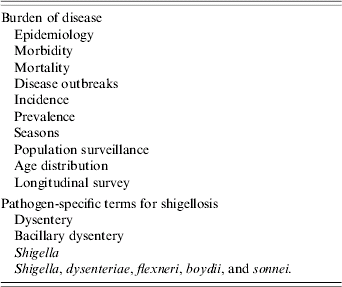
Table 1. Terms used in literature search to identify gaps in data on enteric disease burden
Crude incidence figures are given, without correction for sensitivity of stool culture, or for the proportion of diarrhoeal episodes for which stool specimens were not collected. Publications were sought with information on the incidence of the infection by region according to the 21 regions of the United Nations Department of Social and Economic Affairs, Population Division (http://esa.un.org/unpp/index.asp?panel=5; 2004 revision, accessed 15 January 2007). We examined the correlation between the incidence of Shigella infection and the national per capita gross domestic product (GDP) in year 2000 US dollars, adjusted for purchasing power parity (PPP), at the time that the studies were conducted (http://unstats.un.org/unsd/cdb/cdb_series_xrxx.asp?series_code=29922; accessed 15 January 2007). We searched for publications with information on the age-specific incidence, morbidity, and mortality. We also examined the frequency of Shigella relative to other diarrhoea-causing pathogens including Salmonella, Vibrio cholerae O1, enterotoxigenic Escherichia coli (ETEC), Campylobacter, and rotavirus. In studies that included healthy controls in addition to persons with diarrhoea, we report the frequency of Shigella isolation only among those with diarrhoea. Where stools were tested at two different times of the year, we report data from the period during which a greater number of stool specimens were collected.
RESULTS
Morbidity
Incidence
A 1999 review of published data from 1966 to 1997 estimated the annual global incidence of shigellosis to be approximately 164·7 million episodes, with 163·2 million of those occurring in the developing world [Reference Kotloff4]. These figures were derived by estimating the total number of diarrhoeal cases and multiplying that by the proportion attributable to shigellosis, as determined by the yield of Shigella in stool culture from persons with diarrhoea. While this review distinguished ‘developing’ countries from ‘industrialized’ countries, few studies were available from sub-Saharan Africa or from low-HDI countries, which contributes to uncertainty regarding the true global incidence of shigellosis.
Our review of the published literature from 1984 to 2005 yielded 11 population-based studies of Shigella burden from seven countries (Table 2) [Reference Wang5, Reference Chompook6, Reference Zaki7–Reference Lee16]. Incidence data were reported in six studies and calculated by one of the authors (P. K. R.) based on data provided for five studies. All 11 studies were conducted in medium-HDI countries; no population-based incidence data were available from low-HDI countries. The geographical distribution of shigellosis has been modelled based on available population-based data (Fig. 1). Studies were conducted in Asia (7), Africa (2), and Latin America (2), with both African studies conducted in Egypt. In this limited group of studies, Shigella incidence did not correlate with national GDP (R=−0·11, P=0·75) (data not shown).

Fig. 1. Incidence of shigellosis, by geographic region, 1984–2005. Countries contributing incidence data: China [Reference Wang5], Thailand [Reference Chompook6], Egypt [Reference Zaki7], Bangladesh [Reference Baqui8] and Brazil [Reference Giugliano9].
Table 2. Population-based studies of Shigella incidence published 1984–2005

HDI, Human development index; CFR, case-fatality rate; n.r., not reported.
* Crude incidence reported as no. cases/1000 persons per year.
† Author: incidence reported by author of paper; PKR-calc: incidence calculated by this investigator.
Among the five studies that included surveillance in all age groups, Shigella incidence varied from 0·6 episodes/1000 person-years in Thailand [Reference Chompook6] to 107/1000 person-years in Egypt [Reference Zaki7]. In six studies limited to infants and young children, annual incidence ranged from zero cases (Thai children aged <6 months) [Reference Punyaratabandhu14] to 949 cases/1000 person-years (Bangladeshi children aged <24 months with wasting) [Reference Black10]. Shigella incidence was higher among Egyptian infants aged <6 months (30/1000 child-years) than among infants aged ⩾6 months (23 episodes/1000 child-years) [Reference Zaki7]. In contrast, the incidence among Thai children aged 0–5 months was zero, compared to 86 episodes/1000 person years among children aged 6–11 months [Reference Punyaratabandhu14, Reference Lee16].
Frequency of Shigella relative to other diarrhoeagenic pathogens
Sixty-seven publications reported on the frequency of Shigella and other enteric pathogens in sporadic diarrhoeal illness. Twenty-two studies reported the frequency of Shigella isolation among subjects of all ages (Table 3a), and 45 studies reported on restricted age groups (Table 3b). Data were available from Asia (39 studies), Africa (20), and Latin America (7); one study reported data from four Asian countries [Reference Huilan17].
Table 3a. Relative frequency of endemic Shigella isolation, community- and facility-based studies conducted among all age groups, 1984–2005

HDI, Human development index; Sa, Salmonella (includes typhoidal and non-typhoidal serotypes); ETEC, enterotoxigenic E. coli; VC, Vibrio cholerae; Ca, Campylobacter; RV, Rotavirus; Sh, Shigella; n.t., not tested; n.r., not reported.
* Ranking reported by study author based on identification of other pathogens.
† n.c., Ranking not calculable because testing performed on varying number of samples.
Table 3b. Relative frequency of endemic Shigella isolation, community- and facility-based studies conducted among restricted age groups, 1984–2005

HDI, Human development index; Sa, Salmonella (includes typhoidal and non-typhoidal serotypes); ETEC, enterotoxigenic E. coli; VC, Vibrio cholerae; Ca, Campylobacter; RV, Rotavirus; Sh, Shigella; Comm., community; n.t., not tested; n.r., not reported.
* Ranking reported by study author based on identification of other pathogens.
† Data from four countries reported in a single publication.
Studies varied widely with respect to specific pathogens tested, and diagnostic techniques used. Often, it was not clear whether a given pathogen was tested for and not detected or whether it was not tested for at all. Notably, the frequency of ETEC isolation was reported by 11 of the 20 studies conducted in Africa, 31 out of 40 Asian studies, and four out of seven studies from Latin America. Rotavirus isolation results were reported in 10 (50%) African, 32 (80%) Asian, and four (57%) Latin American studies. Few investigations reported parasitic aetiologies of diarrhoea.
Shigella was isolated from diarrhoeal or dysenteric stools with similar frequency in Asia and Africa (median isolation rate 6%), and with lower frequency in Latin America and the Caribbean (median 3%) (Fig. 2). Shigella was the first or second most frequently isolated pathogen in 10 studies conducted in Africa (n=20), 15 studies in Asia (n=43), and two studies in Latin America (n=7). Rotavirus was tested for in two of the African studies in which Shigella ranked first or second, in six such studies from Asia, and in none of the studies from Latin America and the Caribbean.

Fig. 2. Frequency of Shigella isolation in diarrhoeal or dysenteric stools for all age groups, by geographical area, in studies from medium and low human development index (HDI) countries (n=70 studies).
The isolation rate of Shigella from diarrhoea stools was highest among children aged 1–5 years, with isolation among infants aged <6 months relatively similar to isolation among infants aged 6–12 months (Fig. 3) [Reference Baqui18–Reference Zaman31]. Measles may increase the risk of shigellosis among children, as noted in two papers describing outbreaks of shigellosis following outbreaks of measles [Reference Mathur32, Reference Gupta33].

Fig. 3. Frequency of Shigella isolation in diarrhoeal or dysenteric stools by age group, among children aged <15 years, in studies from medium and low human development index (HDI) countries, 1984–2005 (n=70 studies).
Subgroups
The relative frequency of the four Shigella subgroups in endemic shigellosis was reported by 56 studies (Table 4a). Studies reporting subgroups of fewer than five Shigella isolates were excluded. The median rate of isolation of S. flexneri, S. dysenteriae, S. boydii, and S. sonnei were 57%, 10%, 8%, and 17%, respectively (Fig. 4). S. flexneri was the most commonly detected subgroup in 48 studies and S. dysenteriae was most common in four studies.

Fig. 4. Frequency of Shigella subgroups detected among Shigella isolates from medium and low human development index (HDI) countries, 1984–2005 (n=56 studies).
Table 4a. Frequency of Shigella subgroups in endemic disease, 1984–2005

HDI, Human development index; n.r., not reported; Comm., community.
* Facility type: ‘both’ refers to patient recruitment in both hospital and clinic.
† % dys (S. dysenteriae), % flex (S. flexneri), % boydii, and % sonnei reflect the proportion of the total number of Shigella isolates.
‡ % Sd1 among dys: Sd1 data is presented as % of total Sd1, except where indicated by #, in which case % Sd1 reflects % of all Shigella.
The frequency of S. dysenteriae isolation was inversely correlated with per capita GDP (Fig. 5) (R=−0·54, P<0·0001), and the frequency of S. sonnei isolation was directly correlated with per capita GDP at the time of the study (Fig. 6) (R=0·55, P<0·0001). S. sonnei was the most frequently isolated subgroup in two studies from Thailand published in 2002 and 2005, one from Brazil published in 2005, and one from Turkey published in 2005 [Reference Chompook6, Reference Bodhidatta19, Reference Diniz-Santos21, Reference Ozmert34].

Fig. 5. Frequency of S. dysenteriae among Shigella isolates, by per capita gross domestic product (GDP) (adjusted for purchasing power parity, PPP) of medium and low human development index (HDI) countries, 1984–2005 (n=56 studies) (R=−0·54, P<0·0001). The names of study countries and the years of study are indicated for selected studies.

Fig. 6. Frequency of S. sonnei among Shigella isolates, by per capita gross domestic product (GDP) (adjusted for purchasing power parity, PPP) of medium and low human development index (HDI) countries, 1984–2005 (n=56 studies) (R=0·55, P<0·0001). The names of study countries and the years of study are indicated for selected studies.
None of the population-based studies provide age-specific incidence of Shigella subgroups or serotypes. Among studies reporting isolation rates, three provide data on age-specific isolation of Shigella subgroups [Reference Leano26, Reference MoezArdalan28, Reference Zaman31]. In Bangladesh, S. flexneri was the most frequently isolated subgroup among all age groups except for the 5–9 years group, among whom S. dysenteriae type 1 was more common [Reference Zaman31]. In every age group studied in the Philippines, S. flexneri accounted for at least 75% of Shigella infections including 75% among the 1–2 years age group and 88% among those aged 10–14 years [Reference Leano26]. Based on only 123 total isolates from Iran, S. flexneri was most common among children aged 1–5 years and persons aged ⩾12 years; S. sonnei was most common among children aged <1 year and was as frequent as S. flexneri among children aged 5–12 years [Reference MoezArdalan28].
Fourteen studies assessed the relative frequency of Shigella subgroups during epidemics; 11 focused on persons with dysentery (variably defined as blood with or without mucus in stools) (Table 4b). None of these studies were conducted in Latin America and the Caribbean. Twelve identified a predominance of S. dysenteriae infections, specifically serotype 1, and two identified a predominance of S. flexneri infections.
Table 4b. Frequency of Shigella subgroups in epidemic context, 1984–2005

HDI, Human development index; n.r., not reported.
* Facility type: ‘both’ refers to patient recruitment in both hospital and clinic.
† % dys (S. dysenteriae), % flex (S. flexneri), % boydii, and % sonnei reflect the proportion of the total number of Shigella isolates.
‡ %Sd1 among dys: Sd1 data is presented as % of total Sd1, except where indicated by #, in which case % Sd1 reflects % of all Shigella.
Serotypes
S. dysenteriae is comprised of 15 serotypes, of which type 1 has the capacity to cause large outbreaks [1]. Between 2002 and 2004, numerous publications documented circulating S. dysenteriae type 1 strains resistant to commonly used fluoroquinolones, including ciprofloxacin and ofloxacin [Reference Pazhani35–Reference Dutta41]. The organism remained susceptible only to azithromycin, pivmecillinam, and third-generation cephalosporins.
The World Health Organization (WHO) Collaborating Centre for Shigella at the Centers for Disease Control and Prevention recognizes six serotypes of S. flexneri, which can be further classified into numerous subserotypes, as well as S. flexneri X and Y. Thirteen studies (eight published after 2000), report S. flexneri serotypes in endemic shigellosis (Table 5). Serotype 2a was the most commonly detected serotype in four studies, accounting for 35–67% of S. flexneri isolates; all four studies were conducted in Asia [Reference Chompook6, Reference Bodhidatta19, Reference Dutta42, Reference Varavithya43]. Serotype 1a was the most frequently isolated in a population-based study in China [Reference Wang5]. Serotype 1b was the most frequently detected serotype in Malaysia and the Peruvian Amazon [Reference Jones44, Reference Thong45]. Subserotype 3c was reported from Malaysia and Pakistan, with reports of serotypes 7, 8, and 10 from Pakistan as well [Reference Thong45, Reference Zafar46]. Subserotype 1c has also been reported from Bangladesh and Egypt [Reference El-Gendy47, Reference Talukder48].
Table 5. Frequency of S. flexneri serotypes in endemic setting, 1984–2005

* Number of S. flexneri isolates for which serotype data is provided exceeds number of S. flexneri reportedly isolated in study.
Complications
Few complications were reported in population-based studies of Shigella infection. Dehydration was reported in 11% and 16% of patients [Reference Abu-Elyazeed11, Reference Black12]. Wang et al. reported no cases of rectal prolapse or other gastrointestinal complications in China [Reference Wang5]. Shigella diarrhoea was associated with decreased linear growth rates in a study by Black and colleagues, with the per cent of days of Shigella diarrhoea negatively correlated with linear growth [Reference Black49]. Two studies reported a prolonged duration of diarrhoea with S. flexneri infection compared to infection with other Shigella subgroups [Reference Wang5, Reference Chompook6].
Facility-based studies of shigellosis, albeit probably skewed towards very ill patients and those who can access health-care services, provide additional insight. The rate of hospitalization has ranged from 11% to 47% [Reference Jones44, Reference Khan50–Reference Clemens53]. Dehydration was documented in 2–70% of patients [Reference Hoge52, Reference Rawashdeh54–Reference Thapa56]. Gastrointestinal complications of Shigella infection include persistent diarrhoea (7–38%) [Reference Thapa56–Reference Ahmed59], intestinal obstruction (3–5%) [Reference Thapa56, Reference Bennish60], rectal prolapse (3–38%) [Reference Thapa56, Reference Mudzamiri58], and protein-losing enteropathy (3%) [Reference Chopra61]. The risk of persistent diarrhoea following Shigella infection appears to be increased among young children, in dysenteric episodes of confirmed shigellosis, following infections caused by multiply-resistant Shigellae, or when nalidixic acid (NAL) is administered for infections caused by NAL-resistant organisms [Reference Ahmed59]. Intestinal perforation was reported anecdotally in one study [Reference Grant62].
Haemolytic–uraemic syndrome (HUS), was diagnosed in 1–24% of persons hospitalized with S. dysenteriae type 1 [Reference Mudzamiri58, Reference Chopra61, Reference Al-Qarawi63]. In Bangladesh, HUS was diagnosed in 27% of Shigella-confirmed patients with intestinal obstruction, and none without obstruction [Reference Bennish60]. Three studies reported treatment with antimicrobial agents to which the pathogen is resistant as a risk factor for HUS [Reference Al-Qarawi63–Reference Butler65]. Acute renal failure was documented in 1% [Reference Mudzamiri58] and 25% [Reference Thapa56] of hospitalized patients; neither study specified the proportion of renal failure cases occurring in the context of HUS. Electrolyte imbalances were documented in 42% of patients; both hyponatraemia and hyperkalaemia have been reported [Reference Khan50, Reference Thapa56].
Central nervous system (CNS) manifestations of Shigella infection were noted in 45% of patients in one series [Reference Khan50]. This, and other studies describe complications including seizures (5–27%) [Reference Khan50, Reference Rawashdeh54, Reference Srison and Pornpatkul66] and loss of consciousness (10%) [Reference Khan50, Reference Thapa56]. Factors significantly associated with CNS manifestations include age <15 years, electrolyte imbalance, severe dehydration, fever, shorter duration of illness, higher median weight-for-age, and increased immature leukocytes [Reference Khan50].
Although a recognized complication, this literature search yielded only three reports of reactive arthritis as sequelae to S. flexneri infections [Reference Bhimma64, Reference Butler65, Reference Mazumder67]. Bacteraemia is a relatively rare complication and young age is an important risk factor [Reference Martin68]. Among the handful of adult patients reported to have Shigella bacteraemia, immunocompromising conditions, such as HIV/AIDS are common. One case report from the United States documented recurrent infection with S. boydii in an HIV-infected patient despite 10 days of ceftriaxone therapy, suggesting the need for prolonged therapy in persons with HIV/AIDS and Shigella bacteraemia [Reference Kristjansson69].
Poor nutritional status appears to increase the risk of shigellosis associated with fever, severe dehydration, severe neurological manifestations such as seizures or coma, or requiring hospitalization [Reference Clemens53, Reference Dewan57, Reference Khan70].
Mortality
Only six population-based studies of Shigella incidence included case-fatality rates (CFR) (Table 2). These ranged from 0% [Reference Wang5, Reference Abu-Elyazeed11, Reference Punyaratabandhu14] to 2·6% (Egypt) [Reference Chompook6, Reference Zaki7]. In facility-based studies that reported deaths of hospitalized patients with culture-confirmed endemic Shigella infection, CFRs ranged from 0% to 21% (Table 6).
Table 6. Case-fatality rates in hospital-based studies of endemic shigellosis, 1984–2005

HDI, Human development index; CFR, Case-fatality rate; n.r., not reported; GI, gastrointestinal.
In one study from Bangladesh, patients with shigellosis who were discharged from the hospital (CFR 5%) had a significantly higher risk of post-hospitalization death than persons with watery diarrhoea (CFR 3%) [Reference Bennish and Wojtyniak71]. Reported risk factors for death include young age [Reference Zaman31, Reference Khan50, Reference Thapa56, Reference Bennish72], poor nutritional status [Reference Thapa56, Reference Bennish72, Reference Islam and Shahid73], and CNS manifestations such as altered consciousness or seizures [Reference Khan50, Reference Bennish72]. In a series from North India, 75% of patients who died had renal failure and 25% had Shigella bacteraemia [Reference Thapa56]. Notably, none of these hospital-based studies specifically reported HUS as a risk factor for death. Among severely malnourished children with S. dysenteriae type 1 and S. flexneri, risk factors for death included hypothermia, altered consciousness, low serum blood glucose, and pneumonia [Reference van den Broek74].
A total of 21 studies, all from Africa or Asia, report CFRs of 0% [Reference Niyogi36, Reference Ebright75] to 40% [Reference Pillay76] (median 4%) for epidemic S. dysenteriae type 1 (Table 7). In large epidemics, S. dysenteriae type 1 can account for 0·25–40% of all-cause mortality [Reference Birmingham51, Reference Goma77, Reference Aragon78]. Young children are at increased risk of death from epidemic S. dysenteriae type 1 [Reference Gupta33, Reference Birmingham51, Reference Chopra61, Reference Aragon78–Reference Tuttle83]. Additional risk factors include CNS complications [Reference Sarkar38], malnutrition [Reference Chopra61], the constellation of symptoms and signs associated with HUS [Reference Mudzamiri58, Reference Rollins84], intestinal perforation [Reference Rollins84], and treatment with antimicrobial agents to which the pathogen is resistant [Reference Pillay76].
Table 7. Case-fatality rates in epidemic S. dysenteriae type 1, 1984–2005

HDI, Human development index; CFR, Case-fatality rate; HUS, haemolytic–uraemic syndrome; n.r., not reported.
Temporal distribution
Among 32 studies reporting on seasonal distribution of Shigella infection, 10 were conducted over a period of ⩽1 year and four studies were conducted over a period of ⩾1 year and <2 years. A total of 17 studies were conducted over ⩾2 years, among which seasonality was reported using a variety of terms, including the specific months of the year, the quarters of the year, or more vague terms such as ‘rainy season’ and ‘dry season’. No clear patterns emerged, even when studies were segregated according to northern or southern hemisphere (data not shown).
Pathogen-specific preventive measures
A 2004 review summarizes the current status of Shigella vaccines [Reference Nataro85]. Immunity to Shigella infection, which is directed against the O antigen, is type-specific. Four candidate vaccines using oral formulations (SC 602, CVD 1203, CVD 1204, CVD 1208) and one candidate using the parenteral route (S. flexneri 2a LPS conjugated to recombinant Pseudomonas exoprotein A) target S. flexneri 2a. One oral (WRSS1) and one parenteral vaccine (S. sonnei LPS conjugated to Pseudomonas exoprotein A) target S. sonnei.
Poor toilet and hand hygiene practices play an important role in facilitating Shigella transmission in industrialized and developing countries. Shigellae are also effectively transmitted through contaminated water and survival in water appears to vary by subgroup: S. dysenteriae (2–3 days), S. flexneri (6–47 days), and S. sonnei (35–39 days) [Reference Tuttle83, Reference Iwamoto86–Reference Mitscherlich89]. Foods, particularly contaminated produce, are well-recognized as vehicles for Shigella transmission [Reference Hoge52, Reference Tuttle83, Reference Naimi90]. Flies have been implicated and may be especially important in settings where latrines are in close proximity to food preparation or consumption areas [Reference Levine and Levine91–Reference Househam94]. Promotion of handwashing with soap has been linked to reduced Shigella transmission in endemic and epidemic settings [Reference Khan95, Reference Mohle-Boetani96]. Specific latrine behaviour may be important. Anal cleansing rags used communally were implicated in one outbreak of S. dysenteriae type 1 [Reference Birmingham51]. To the extent that the use of toilet paper reduces hand contamination with stool, this simple intervention may also be useful for reducing Shigella transmission [Reference Aung Myo97]. Notably, one study has demonstrated dramatic reductions in Shigella incidence with aggressive measures to control houseflies, which may breed in latrines [Reference Cohen92].
Diagnostics
Culture of whole stools or stool collected by rectal swab is the most widely used technique for diagnosis of Shigella infection. However, Shigellae are relatively fragile organisms compared with other enteric pathogens. Yield from stool culture is increased when the specimen is placed in transport medium and held at refrigeration temperatures (4°C) if not cultured immediately [98]. Buffered glycerol saline is the preferred medium but Cary–Blair medium is also acceptable. Polymerase chain reaction (PCR) techniques have been used to identify Shigella genes coding for the invasion plasmid antigen H (ipaH), which is present in all Shigellae and in enteroinvasive E. coli, and Shiga toxin (stx), which is present in S. dysenteriae type 1 and in enterohaemorrhagic E. coli. When compared with PCR testing for Shigella primers, the sensitivity of culture has been estimated at 72% [Reference Islam99].
Recent data suggest that an innovative transport technique (DNA/RNA Protect™, Sierra Diagnostics Inc., Sonora, CA, USA) permits detection of ipaH from stool specimens held for prolonged periods at room temperature [Reference Hyytiä-Trees100]. An enzyme-linked immunosorbent assay (ELISA) is available for detection of Shiga toxin 1. The ELISA was evaluated in a single study in Bangladesh, and demonstrated 95% sensitivity and 85% specificity for detection of S. dysenteriae type 1 [Reference Salam101].
DISCUSSION
This review highlights the large gaps in data on the burden of Shigella infections for low-HDI countries and, more specifically, for sub-Saharan Africa (Table 8). It also identifies additional research needed with respect to the burden of disease among infants and population-based rates of Shigella-associated complications and mortality. Comprehensive microbiological, clinical, and epidemiological studies using newer and more sensitive diagnostic methods to determine the relative importance of Shigella and other diarrhoeal pathogens could help address these data gaps and further inform prevention efforts.
Table 8. Data gaps and research needs for Shigella infections in medium and low human development index (HDI) countries

The absence of population-based incidence data from sub-Saharan Africa and from low-HDI countries is our most striking finding. Because of their low development status, and limited access to improved water supplies and sanitation facilities, these countries may be expected to have a higher burden of Shigella infections than the medium-HDI countries where population-based studies have been conducted. Moreover, given the additional lack of substantial data on shigellosis in the context of HIV disease, active population-based surveillance studies conducted in this important geographic region to elucidate overall disease burden, frequency of subgroups, serotypes, and subserotypes, are critical for understanding the true global burden of Shigella infections.
This review detected wide variation in the incidence of Shigella infections from the few available population-based studies, ranging from 0 episodes/1000 person-years [Reference Punyaratabandhu14] to 949 episodes/1000 person-years [Reference Black10]. However, comparisons between these studies is challenging because of variations in the age groups under study, methodologies for surveillance, and techniques for specimen collection, transport, and culture.
Several population-based studies included only young children; others, which included all age groups, reported incidence data only by age group and not for the population as a whole. Where possible, future population-based studies should describe age group-specific and total incidence, and include isolation rates for all Shigella and for the specific subgroups and serotypes.
The risk of shigellosis among young infants is poorly characterized. This review identified very few studies that separated out infants aged <6 months who are likely to be breastfeeding and, thus, exposed to maternal IgA [Reference Hayani102]. Population-based data on the incidence of Shigella infections among infants aged <6 months and 6–11 months could help inform this discussion, which is critical to the development and deployment of Shigella vaccines.
Our attempts to estimate the frequency of Shigella relative to other diarrhoeal pathogens revealed important variations in the spectrum of pathogens sought. Rotavirus, the most common cause of severe diarrhoeal illness worldwide [Reference Parashar103], was not tested for in a majority of the ten African studies that found Shigella to be the first or second leading cause of diarrhoea. This review underscores the need for comprehensive stool analyses, including identification of Rotavirus, the different groups of diarrhoeagenic E. coli, and parasitic infections, in aetiological studies of diarrhoeal disease. Moreover, such studies are particularly needed in sub-Saharan Africa.
A vast amount of data has been published between 1984 and 2005 regarding the relative frequency of the four Shigella subgroups. The predominance of S. flexneri above other subgroups, particularly in endemic shigellosis, is clear. S. sonnei remains the most frequently isolated subgroup in industrialized countries [Reference Kotloff4]. Even among medium- and low-HDI countries, there is a significant correlation between the GDP of the country at the time of the study and the frequency of S. sonnei isolation. The predominance of S. sonnei over S. flexneri in recent studies from Thailand, Brazil, and Turkey probably reflects the expanding economies in these three countries.
Given their respective importance to epidemics and frequency of isolation, we focused this review on S. dysenteriae serotype 1 and on the serotypes of S. flexneri. Large epidemics of S. dysenteriae type 1 were documented in Central America in the 1960s and 1970s, across Africa during the 1980s and 1990s and in South Asia during the early part of each decade between 1970 and the present [Reference Reller104–Reference Wittenberg106]. Studies reporting on the frequency of Shigella subgroups in the epidemic context indicate that during an epidemic period, a single subgroup, particularly S. dysenteriae serotype 1, can explain the majority of Shigella infections [Reference Goma77]. With each reemergence, S. dysenteriae type 1 has exhibited an expanding resistance profile. Most recently, strains of S. dysenteriae type 1 circulating in South Asia have demonstrated resistance to multiple fluoroquinolones, with susceptibility only to azithromycin, pivmecillinam and third-generation cephalosporins [Reference Pazhani35–Reference Dutta42Reference Pazhani35–Reference Dutta42, Reference Jahan and Hossain105]. Studies of the efficacy of these agents against S. dysenteriae type 1 are rare and have only been conducted in adults [Reference Kabir107, Reference Kabir108]. It is vital to better characterize effective antimicrobial agents and regimens for the treatment of multiply-resistant S. dysenteriae type 1 infection in children and adults ahead of the next epidemic. Indeed, multi-drug resistance (MDR) is a common feature of the other Shigella subgroups as well. Identifying the best therapeutic strategies for MDR Shigella remains a central research priority in the study of shigellosis.
Information on the subserotypes of S. flexneri is relevant because of the implications for vaccine development; most recent studies identified in this review provided that information. Currently, subserotypes 1c and 3c, and serotypes 7, 8, and 10, are not recognized by the WHO Collaborating Centre for Shigella at the Centers for Disease Control and Prevention (CDC). The International Committee on Systematic Bacteriology Subcommittee on the Taxonomy of Enterobacteriaceae recognized only two subserotypes within S. flexneri serotype 1 (1a and 1b) and serotype 3 (3a and 3b) in 1984 [Reference Brenner109]. It is important to confirm the identification of serotypes 7, 8, and 10 reported from Pakistan and to assess their contribution to S. flexneri disease elsewhere.
A large number (18%) of untypable S. flexneri isolates was reported from Pakistan, a finding also noted in studies from Vietnam and Bangladesh, in which 40% and 12% of S. flexneri, respectively, could not be definitively serotyped [Reference Isenbarger15, Reference Zafar46, Reference Talukder110]. Several studies reported the serotype of <100% of S. flexneri infections [Reference Chompook6, Reference El Nageh23, Reference Jones44, Reference Donald111], suggesting that some isolates could not be definitively serotyped. The prevalence of untypable S. flexneri isolates in geographical regions other than South and South East Asia, and the implications of these findings for vaccine development demand further study.
Reliable typing antisera for S. flexneri are not easily produced because of the need to absorb cross-reacting antibodies. In the past, quality assurance and quality control problems have been noted in commercially available typing antisera [Reference Evins112]. Improved quality assurance and quality control of commercially distributed typing antisera is necessary to better define the epidemiology of S. flexneri.
The literature search strategy yielded many reports about the complications of Shigella infection; however, most of these were from hospital-based studies that are biased towards the most severely ill patients with shigellosis. An important gap in the literature is the absence of population-based rates of Shigella complications. Such surveillance may require longer-term follow-up of larger populations than most demographic surveillance systems currently conduct.
Our review yielded Shigella mortality data from six population-based studies. None of the five population-based studies conducted before 1990 provided mortality information. Calculation of mortality rates in population-based studies are critical for estimating the true public health impact of shigellosis; however, such studies are rightfully conducted under strict human subjects research guidelines, mandating prompt and appropriate treatment for bloody diarrhoea. Thus, it is difficult to study the natural history of Shigella complications or mortality in such a research setting. Although unbiased estimates of Shigella mortality may be difficult to obtain, the consistent use of verbal autopsy data and molecular diagnostic techniques in population-based surveillance may help. Risk factors for death in endemic shigellosis were available only from papers from South Central Asia and from one study in Jordan. Identification of modifiable risk factors for death from all regions and strategies for prevention of these risk factors are important needs in the study of Shigella.
Despite a number of studies reporting on the temporal distribution of Shigella infections, no clear patterns emerged with respect to seasonality. The use of non-standard terminology and data collection for <2 consecutive years make comparisons of seasonal trends across studies difficult. We recommend the collection and analysis of temporal distribution data for shigellosis over multi-year periods, using consistent descriptors and contextual environmental information such as rainfall and flooding.
Since Shigellae, like many other enteric pathogens, are transmitted through consumption of contaminated food and drinking water, improvements in sanitation, drinking water quality, and food preparation and storage practices in homes and in communities are expected to have dramatic impacts on the burden of shigellosis. Because the pathogen has a low infectious dose, the promotion of handwashing with soap after defecation would be expected to reduce the risk of Shigella transmission [1]. However, specific data regarding the beneficial effects and feasibility of scaling up of household-level control measures, such as promotion of handwashing with soap, improved latrine design to include handwashing stations, aggressive fly control, and modification of culturally ingrained anal cleansing habits are largely lacking.
A number of candidate Shigella vaccines are under development. Oral formulations would probably be most useful for the developing world. In September 2006, the International Vaccine Institute, with partners in various Asian countries, estimated the burden of Shigella infections and identified the frequency of subgroups and serotypes. Their work demonstrates that 90% of S. flexneri infections are caused by eight different serotypes and subserotypes [Reference Von Seidlein113, Reference Sansonetti114]. This diversity suggests that the identification of an antigen common to most or all of the S. flexneri serotypes would facilitate vaccine development significantly. While extensive attention has been paid to the development of Shigella vaccines, and data has been gathered regarding Shigella burden in Asia, relatively little information is available regarding the epidemiology of shigellosis and, importantly, the feasibility of introducing these vaccines in sub-Saharan Africa.
Finally, any estimation of the burden of Shigella infections must take into account the limitations of currently utilized specimen transport and diagnostic methods [98]. Thus, even the culture-based estimates of Shigella incidence reported above may underestimate the true burden of disease. Improved transport methods, such as the DNA/RNA Protect™ specimen collection system, must be validated for the detection of Shigella in clinical settings, especially in remote areas. Population-based surveillance for Shigella incidence using potentially more sensitive methods, such as PCR techniques, in addition to culture, may improve Shigella incidence estimates [Reference Vu115]. Moreover, development and evaluation of rapid gene-based diagnostic techniques for faecal specimen testing, available to the serotype level, may enhance measurement of the burden of Shigella and its various serotypes. However, given their potential for high sensitivity, these methods should be evaluated to understand the extent to which they detect clinically relevant infections vs. asymptomatic carriage.
In summary, this paper describes the acute need for population-based studies of Shigella morbidity and mortality from sub-Saharan Africa, and from low-HDI countries in general. Such information would be invaluable for the prioritization of precious public health resources for pathogen-specific interventions, such as vaccination programmes targeting Shigella. Additional data regarding complications, modifiable risk factors for mortality, the burden of disease among infants, effectiveness of non-vaccine prevention measures, and improved diagnostics would add greatly to our knowledge of Shigella infections.
ACKNOWLEDGEMENTS
This work was supported in part by the U.S. National Institutes of Health Fogarty International Center and by grant number 32143 from the Bill and Melinda Gates Foundation ‘Assessment of diarrhea disease burden and public health programs to control diarrhea in Asian subcontinent and Africa’.
DECLARATION OF INTEREST
None.


