


MC twins are at substantially increased risk of adverse outcome compared to DC twins (Sebire et al., Reference Sebire, Snijders, Hughes, Sepulveda and Nicolaides1997). This excess of adversity in MC twins is mainly attributed to the complications resulting from connected circulation (Lewi et al., Reference Lewi, Jani, Blickstein, Huber, Gucciardo, Van Mieghem and Deprest2008). The vascular anastomoses are the anatomical basis for connected circulation within twin pairs. Three types of vascular anastomoses are reported in injection studies of MC placentas, namely arterio-arterial (AA) anastomoses, veno-venous (VV) anastomoses, and arterio-venous (AV) anastomoses. The unidirectional blood flow in AV anastomoses enables volume disequilibrium, resulting in severe complications such as TTTS and TAPS (Lopriore et al., Reference Lopriore, van den Wijngaard, Middeldorp, Oepkes, Walther, van Gemert and Vandenbussche2007). The association between TTTS/TAPS and vascular anastomoses in MC twins has been extensively illustrated in placental injection studies (De Paepe et al., Reference De Paepe, Shapiro, Greco, Luks, Abellar, Luks and Luks2010; de Villiers et al., Reference de Villiers, Slaghekke, Middeldorp, Walther, Oepkes and Lopriore2013; Reference de Villiers, Zhao, Cohen, van Zwet, Duan, Oepkes and Lopriore2015; Denbow et al., Reference Denbow, Cox, Taylor, Hammal and Fisk2000; Zhao et al., Reference Zhao, Cohen, Middeldorp, Klumper, Haak, Oepkes and Lopriore2014). In contrast, little is known of the vascular anastomoses in DC placentas due to lack of placental injection for DC placentas. In addition, placental share discordance is quite common in MC twins, leading to discordant fetal growth, even selective intrauterine growth restriction (sIUGR; Fick et al., Reference Fick, Feldstein, Norton, Wassel Fyr, Caughey and Machin2006; Lewi et al., Reference Lewi, Cannie, Blickstein, Jani, Huber, Hecher and Deprest2007). Again, since placental injection is not routine practice for the examination of DC placenta, the placental share discordance in DC twins remains to be elucidated. The aim of this study was to compare the placental characteristics between a large cohort of MC and DC placentas using colored dye injection.
Materials and Methods
All placentas of twin pregnancies consecutively delivered at Leiden University Medical Center (the Netherlands) and Medical University of Warsaw (Poland) from September 2012 to December 2015 were eligible for this study. MC placentas treated with fetoscopic laser surgery were excluded. We also excluded twin placentas with single or double fetal demise, incomplete injection due to maceration, fixation in formalin, and severe damage. Chorionicity was evaluated during the 11–14 weeks’ sonographic examination and was confirmed post-natally by macroscopic or microscopic histopathological evaluation. The type of umbilical cord insertion and number of umbilical vessels were recorded. Velamentous cord insertion was defined as the insertion of umbilical cord into the amniotic membrane instead of the placental parenchyma. All twin placentas were injected according to the protocol published previously (Lopriore et al., Reference Lopriore, Slaghekke, Middeldorp, Klumper, van Lith, Walther and Oepkes2011). After injection, the type and number of vascular anastomoses were documented. Digital placental pictures were taken for various further computerized analysis, such as measurement of placental share and anastomostic size. Individual placental share was measured as the venous return area of each twin using Image J 1.45s (Image J, National Institute of Health, USA). Placental share difference was calculated as the larger placental share minus the smaller placental share. Placental share discordance was calculated using the following formula: (larger placental share — smaller placental share)/larger placental share × 100%. Part of the placental data were reported to describe a special type of AA and VV anastomoses, the so-called partially hidden AA and VV anastomoses (Zhao et al., Reference Zhao, Dang, Haak, Middeldorp, Klumper, Oepkes and Lopriore2015).
The following perinatal variables were collected prospectively: TTTS, TAPS, sIUGR, gestational age at birth, birth weight, Hb levels at birth, and delivery mode. Diagnosis of TTTS was based on the Eurofetus criteria (Senat et al., Reference Senat, Deprest, Boulvain, Paupe, Winer and Ville2004). TAPS was defined as the diagnostic criteria proposed by Slaghekke et al. (Reference Slaghekke, Kist, Oepkes, Pasman, Middeldorp, Klumper and Lopriore2010). Birth weight discordance was calculated by the following formula: (larger twin — smaller twin)/larger twin × 100%. sIUGR was defined as a birth weight discordance of ≥25% (Lopriore et al., Reference Lopriore, Pasman, Klumper, Middeldorp, Walther and Oepkes2012). Individual birth weight share was calculated by dividing the birth weight of each infant by the sum of the birth weights of both infants. Birth weight share/placental share ratio was calculated by dividing the birth weight share by the corresponding placental share (Lewi et al., Reference Lewi, Cannie, Blickstein, Jani, Huber, Hecher and Deprest2007; Zhao et al., Reference Zhao, Slaghekke, Middeldorp, Duan, Oepkes and Lopriore2014).
Statistics
The Kolmogorov–Smirnov test was adopted to assess the normality of continuous variables. Data were analyzed using chi-square, Fisher exact, Mann–Whitney or Student t-tests, as appropriate. Spearman r was generated to evaluate the correlation between placental share and birth weight share. Statistical significance was considered if a p value was less than .05. Data were analyzed using GraphPad Prism v6.0 (GraphPad Software Inc. La Jolla, CA 92037 USA) and IBM SPSS Statistics 22.0® (IBM Corporation, Armonk, New York, USA).
Results
A cohort of 267 eligible twin placentas were examined at both centers during the study period, including 143 MC placentas and 124 DC placentas. Nine (3%) placentas were excluded due to incomplete injection. The remaining 134 MC placentas and 124 DC placentas were analyzed in this study. In the group of MC twins, 18 (13%) were complicated with TTTS (not treated with fetoscopic laser surgery), and there were 8 cases (6%) with TAPS and 31 (23%) cases with growth discordance. Neither TTTS nor TAPS occurred in the group of DC twins, whereas growth discordance occurred in 10% (12/124) of DC twins. Additional characteristics of the two groups are shown in Table 1.
TABLE 1 Baseline Characteristics
Data was displayed as mean ± SD, median (IQR) or n (%).
Vascular anastomoses were detected in 99% (133/134) MC placentas and 0% (0/124) DC placentas, respectively (p < .01). In the group of MC placentas, the frequency of AV anastomoses, AA anastomoses, and VV anastomoses was, respectively, 99% (133/134), 85% (114/134), and 28% (38/134). The median number of vascular anastomoses per MC placenta was 11 (interquartile 6–18). One percent (1/134) of MC placentas consisted of two separate placental masses (so-called bipartite MC placentas). Forty-four percent (54/124) of DC placentas had two separate placental masses, whereas the rest of DC placentas were fused. Comparisons of placental characteristics between MC and DC placentas are summarized in Table 2. Examples of MC and DC placentas after colored dye injection are illustrated in Figures 1 and 2, respectively.
TABLE 2 Comparison of Placental Angio–Architecture Between MC and DC Placentas
Data were displayed as median (IQR) or n (%). aDenotes the presence of velamentous cord insertion per infant instead of twin pair. bValue was given as median (95% CI).

FIGURE 1 A monochorionic placenta after colored dye injection. The blue, white and yellow arrows indicate the AA anastomoses, VV anastomosis and AV anastomoses, respectively. The white-dotted line indicates the vascular equator. The first twin had a placental share of 67% and the second twin 33%.
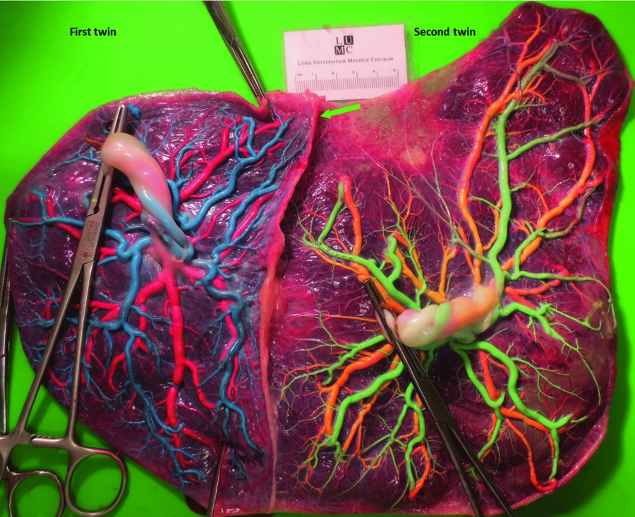
FIGURE 2 A dichorionic placenta after colored dye injection. The two placental masses were fused. No vascular anastomoses were detected after injection. The green arrow indicates the inter-twin septum. The individual placental share in first and second twin was 39% and 61%.
We further related the individual placental share to birth weight share in MC and DC twins to evaluate the relation between fetal growth and placenta share. We found that birth weight was significantly associated with placental share in both MC twins (Spearman r = 0.64, 95% confidence interval 0.56–0.71, p < .01, Figure 3) and DC twins (Spearman r = 0.32, 95% confidence interval 0.19–0.44, p < .01, Figure 3).

FIGURE 3 Correlation between placental share and birth weight in MC twins (Spearman r = 0.64; 95% CI: 0.56–0.71; p < .01) and DC twins (Spearman r = 0.32; 95% CI: 0.19–0.44; p < .01).
Discussion
This is the first study to compare the angioarchitecture between MC and DC placentas using an accurate and reliable technique. We found that vascular anastomoses are almost always present in MC placentas but non-existent in DC placentas. As a result, hematological and perinatal complications due to shared circulation by vascular anastomoses occur only in MC twins, but not in DC twins.
The vascular anastomoses and associated consequences in MC twins have been well studied. In accordance with previous placental injection studies, this study shows that the presence of vascular anastomoses in MC placentas is quite common (De Paepe et al., Reference De Paepe, Burke, Luks, Pinar and Singer2002; Lewi et al., Reference Lewi, Cannie, Blickstein, Jani, Huber, Hecher and Deprest2007; Robertson & Neer, Reference Robertson and Neer1983; Zhao et al., Reference Zhao, de Villiers, Slaghekke, Walther, Middeldorp, Oepkes and Lopriore2013). In contrast, the presence of vascular anastomoses in DC placentas has not systematically been studied with colored dye injection. However, several case reports have reported on DC twins with placental vascular anastomoses (Biran et al., Reference Biran, Bornes, Aboura, Masmoudi, Drunat, Baumann and Baud2011; French et al., Reference French, Bieber, Bing and Genest1998; Lage, Reference Lage, Vanmarter and Mikhail1989; Phelan et al., Reference Phelan, Geer and Blackburn1998; Quintero et al., Reference Quintero, Kontopoulos, Barness, Steffensen, Hilbelink, Chmait and Bornick2010; Rodriguez et al., Reference Rodriguez, Porter, Stirrat and Soothill1996). In these reports, vascular anastomoses were inspected when associated complications were suspected, such as TTTS, TAPS, and twin reversed arterial perfusion (TRAP). Nevertheless, Robertson and Neer (Reference Robertson and Neer1983) reported a paucity of vascular anastomoses in DC placentas with a fused mass. Thus, the vascular anastomoses in the general population of DC twins remains uncertain. In this study, we consecutively examined a large cohort of DC placentas with colored dye injection and did not detect any vascular anastomoses. This disparity in vascular anastomoses between MC and DC placentas may be due to the distinct embryological process. In DC twins, a pre-requisite for the formation of vascular anastomoses is that the chorionic vessels of one twin pass through the chorion and amnion of both twins into the placental territory of the co-twin. This process may be not only hampered by mechanical factors, but also be inhibited by the chemical factors in amnion (Niknejad et al., Reference Niknejad, Paeini-Vayghan, Tehrani, Khayat-Khoei and Peirovi2013).
In this study, we found that birth weight was strongly associated with placental share in both MC and DC twins. Our findings support the theory proposed by Salafia et al. (Reference Salafia, Kiryankova, Inany, Charlagorla, Park, Khawar and Lederman2016) that the growth relationship between birth weight and placental weight is comparable between MC twins and DC twins. Interestingly, unequal placental share appears to be less frequent in DC twins than MC twins despite the common existence of inter-twin competition for space and nutrition in both types of twins. Several studies argue that the blastocyst allocated to each twin is disequilibrated during the twining process of MC twins, leading to different growth potential within twin pairs (Silva et al., Reference Silva, Martins, Matias and Blickstein2011). In addition, implantation into an unfavorable milieu of one twin may also play a role in the increased frequency of unequal placental share in MC twins, given the higher prevalence of velamentous cord insertion indicative of insufficient placentation (Costa-Castro et al., Reference Costa-Castro, De Villiers, Montenegro, Severo, Oepkes, Matias and Lopriore2013).
This study has several limitations. One is the selection bias due to the referral nature of our centers. Twin pregnancies referred to our centers usually undergo a complicated course, especially MC twins. Since vascular anastomoses and unequal placental share are significantly related to the adverse outcome in MC twins (Lewi et al., Reference Lewi, Deprest and Hecher2013), the findings on MC twins in this study may be overestimated. However, the prevalence of TTTS, TAPS, and sIUGR detected in the MC twin cohort in this study is comparable to the expected prevalence in an unselected cohort of MC twins (Lewi et al., Reference Lewi, Jani, Blickstein, Huber, Gucciardo, Van Mieghem and Deprest2008). Another possible limitation is that individual placental share in DC twins may not represent the size of individual placental mass. Placentometric studies show that many aspects of placental gross morphology are associated with fetal growth, including area of placental surface and placental weight (Barker & Thornburg, Reference Barker and Thornburg2013). Unfortunately, the weight of individual placental mass was not measured in this study. Finally, minuscule vascular anastomoses have also been discovered underneath the placental surface using a casting technique with latex injection. In this study, placental casting was not performed and the presence of deep-hidden anastomoses was not evaluated (van den Wijngaard et al., Reference van den Wijngaard, Lopriore, van der Salm, Schaap, Vandenbussche, Deruiter and van Gemert2007).
In conclusion, vascular anastomoses are extremely rare (and almost non-existent) in DC placentas, but ubiquitous in MC placentas. In addition, unequal placental sharing appears to occur more frequently in MC twin placentas. The two placental characteristics are responsible for the increased risk of perinatal complications associated with MC twinning.


