


Case History
A 42-year-old woman presented to the emergency department (ED) with acute onset periumbilical pain and erythema, which started that morning. She felt nauseated but denied vomiting. Her bowel movements were regular and non-bloody. She did not have a fever. She had no significant prior medical history, no recent trauma, and had no previous surgeries.
In the ED, she appeared uncomfortable from pain but was not in acute distress. Vital signs upon initial presentation included a heart rate of 120 beats/min, a blood pressure of 145/111 mm Hg, respiratory rate of 16 breaths/min, temperature of 36.8°C (98.2°F), and an oxygen saturation of 99% on room air. Her vital signs two hours later after a fluid challenge and analgesics had improved to a heart rate of 74 beats/min and blood pressure of 132/83 mm Hg, and the remaining vitals were the same. Her abdomen demonstrated periumbilical erythema, with a central clearing; the area was tender to palpation, and warm to touch. Images of her periumbilical region are shown in Figure 1. The rest of the abdomen was soft and non-tender, with no masses or organomegaly. An ED ultrasound was performed to assess for possible subcutaneous abscess, which was not seen. Cardiovascular and respiratory exams were unremarkable.
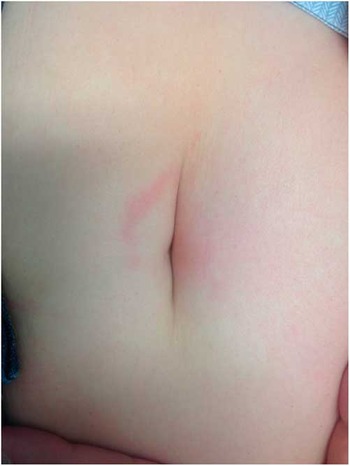
Figure 1 Periumbilical rash with central clearing.
Question
The correct diagnosis is:
-
a) Omphalitis
-
b) Cellulitis
-
c) Lyme disease
-
d) Subcutaneous abscess
-
e) Erythema multiforme
For the answer to this challenge, see next page.
Answer
The correct diagnosis is c. Figure 1 demonstrates the characteristic erythema migrans rash, pathognomonic of Lyme disease. Of interest, the patient initially had a CT of the abdomen ordered to rule out intra-abdominal pathology until a tick was found in the patient’s umbilicus and removed intact, changing the ED approach (Figure 2). Appropriate Lyme disease serology was sent, and the tick was analyzed by public health laboratories which demonstrated an Ixodes scapularis, also known as a “deer tick” or “black-legged tick.” The patient was diagnosed with early localized Lyme disease, given that she showed no features of disseminated infection. She was sent home on a 21-day course of doxycycline without awaiting serologic results.

Figure 2 Black-legged tick removed intact from umbilicus.
Discussion
Lyme disease is caused by the spirochete Borrelia burgdorferi; this is generally spread through the bite of a black-legged tick. B. burgdorferi begins to replicate in the skin and proceeds to spread through the bloodstream, causing overlapping stages of the disease. These stages include early localized disease, early disseminated infection and late disease.Reference Shin 1 Clinical findings in early localized Lyme disease are variable, but commonly include: a characteristic expanding erythema migrans rash and some combination of constitutional symptoms. One challenge in diagnosis is the variable presentation of erythema migrans. It is seen in 80% of patients, ranging anywhere from 1-36 days after the bite. The colour varies from pale red to dark purple, and, while often flat, can have raised elements. The classic description includes a central clearing or “bull’s-eye” appearance, but may be homogenous. While most lesions occur at the site of the tick bite, approximately 20% of patients will have multiple lesions develop.Reference Tibbles and Edlow 2
Early disseminated disease can last for several months. Presentations may include neurological findings such as Bell palsy, radiculopathies and meningitis. In addition carditis and large joint arthritis can be seen. Finally, late Lyme disease can last months to years, with various rheumatologic and neurologic manifestations throughout the body.Reference Shapiro 3 - Reference Lindsay, Bernat and Dibernardo 6
Diagnosis of Lyme disease is made clinically in early localized Lyme disease, often based on the presentation of an erythema migrans rash. If a clinical diagnosis is made, treatment should be initiated immediately. Serologic testing may be performed, but should not prevent initiation of treatment, and may be negative in early disease. Diagnostic testing is generally reserved for patients with symptoms of disseminated infection, who have had exposure to black-legged ticks.Reference Lindsay, Bernat and Dibernardo 6 This is done through a two-tiered serologic testing method, including a screening ELISA test, which detects the presence of IgM and IgG antibodies to B. burgdorferi. This is followed up by a Western blot, if results from the screening test are positive. 7 It is also important to note that a positive serologic test does not distinguish between a current and past infection, as antibodies will remain detectable.Reference Wormser, Levin and Soman 8
Ticks removed from the skin can also be sent in to the Public Health Agency of Canada for further surveillance of the distribution of the tick population. 7 When removing a tick from the skin, tweezers should be used to grasp the tick close to the skin. The tick should be pulled upward slowly, and careful attention should be made to ensure the tick does not get twisted or crushed. Patient counseling on the signs and symptoms of early Lyme disease should be provided.Reference Wesolowski, Wozniak and Mila-Kierzenkowska 9 Chemoprophylaxis can be considered, with a single dose of doxycycline (200 mg), for patients who have had a tick attached to them for more than 24 hours.Reference Hachette, Davis and Johnson 4
In Canada, Lyme-endemic areas are found in suitable woodland habitats for ticks, generally the southern parts of each province, with a flare-up of cases from late spring to early fall.Reference Moore, Eisen and Monaghan 10 Ongoing surveillance is continually done through the Public Health Agency of Canada, as Lyme disease is a nationally notifiable disease. 7 The physician should have knowledge of local tick populations, as Lyme disease should be included on the differential diagnosis for those patients at risk.
Treatment for adults with Lyme disease is doxycycline, 100 mg orally twice daily for 14-21 days. Other treatment regimens include cefuroxime or amoxicillin, depending on the patient’s age, pregnancy status, allergies and medical comorbidities.Reference Ogden, Artsob and Lindsay 5 This varies drastically from the treatment of cellulitis, which is mainly cephalexin or cloxacillin, These antibiotics may be of little value in the treatment of Lyme disease, and therefore keeping it in the differential, and using appropriate treatment with doxycycline, is pivotal in preventing disseminated disease.
Overall, physical findings, including the classic erythema migrans rash, may suggest the diagnosis of Lyme disease, and treatment should be started without waiting for serologic confirmation. As this case illustrates, physical findings can present in unusual places, therefore the physician must maintain a high level of suspicion for Lyme disease in endemic areas.
Competing Interests: None declared.
Keywords: Bedside Ultrasound, Emergency Department, Lyme Disease, Rash, Erythema Migrans


