


- DR
-
diurnal rhythm
- FGF-23
-
fibroblast growth factor-23
- PTH
-
parathyroid hormone
- uCa/uCr
-
urinary Ca/urinary creatinine
- uP
-
urinary P
Ethnicity is generally defined as ‘the fact or sense of belonging to a particular ethnic group’( 1 ). An ethnic group shares a distinctive cultural and historical tradition, often associated with nationality, or region by which the group identifies itself and others recognise it( 1 ). This broad concept of ethnicity, encapsulating culture, lifestyle, identification, environment, biology and genetics, is what we refer to in this review.
Ethnic differences in bone health
The prevalence of osteoporosis and the incidence of age-related fragility fracture vary by ethnicity. Kanis et al. report a greater than 10-fold variation in fracture probability between countries across the world( Reference Kanis, Oden and McCloskey 2 ), and substantial differences are also seen between ethnic groups within a country. The age-adjusted incidence rates of hip and distal-forearm fractures in elderly West African men and women are shown to be substantially lower than those found in Britain( Reference Adebajo, Cooper and Evans 3 ). Similarly, Yan et al. have reported a low incidence of hip fracture in Northern China, and the overall male-to-female ratio for hip fracture incidence was 1·15:1 in contrast with that of 0·34:1–0·43:1 in populations of most Western countries( Reference Yan, Zhou and Prentice 4 ). Differences are also found within the USA between ethnic groups. Age-adjusted hip fracture rates were reported to be 57/100 000 in African-American females, compared with 140/100 000 in White-American females( Reference Silverman and Madison 5 ), with similar trends for men( Reference Barrett, Baron and Karagas 6 ). Also, Asian and Hispanic men and women living in the USA have lower hip fracture rates than their White-American counterparts( Reference Silverman and Madison 5 , Reference Kellie and Brody 7 , Reference Lauderdale, Jacobsen and Furner 8 ). It appears that across the world, White populations are consistently reported to be at a higher risk of fracture than other ethnic groups( Reference Kanis, Oden and McCloskey 2 – Reference Yan, Zhou and Prentice 4 , Reference Kellie and Brody 7 , Reference Lauderdale, Jacobsen and Furner 8 ). These differences cannot be fully explained by a lower bone mass in White populations. While African-Americans tend to have a higher bone mineral density than White-Americans, this is not the case for Asian-Americans, Asians or Africans. Fracture risk is multi-factorial and only partly depends on bone mass. Other factors influencing fracture risk are likely to be involved in the observed ethnic differences in bone health.
The multitude of factors determining fracture risk can be subdivided into extra-skeletal and skeletal factors. The former include age, gender, neuromuscular coordination, falls, and in the case of a fall, adiposity and soft tissue cushioning may absorb the force and thus influence the impact on the bone. Skeletal factors include bone mass, geometry, the rate of bone turnover and mineral metabolism. Mineral and bone metabolisms are intimately interlinked and this review focuses on ethnic variation in the metabolism of the bone forming minerals, Ca and phosphate (P).
Ca and P metabolism
Bone consists of an extracellular mineralised matrix consisting of mineral salts (mainly hydroxyapatite (Ca10(PO4)6(OH)2) crystals) bound to proteins. The metabolic function of bone is crucial for Ca and P homoeostasis. Ca, P and bone metabolism are regulated by common factors, mainly parathyroid hormone (PTH), 1,25-dihydroxy vitamin D (1,25(OH)2D) and fibroblast growth factor-23 (FGF-23). The parathyroid gland detects the fluctuations in plasma ionised Ca (Ca2+) concentration through the Ca2+-sensing receptor. In response to a reduction in plasma Ca2+, PTH is secreted by the parathyroid cells. In the kidney, PTH binds to the PTH receptor and stimulates renal Ca reabsorption from the urinary filtrate( Reference Favus, Goltzman and Rosen 9 ). This occurs through a multitude of actions, including the increase of the renal Na/K/Cl2 co-transporter to stimulate paracellular Ca reabsorption, and through the transient receptor potential channel TRPV5, to stimulate luminal Ca transfer( Reference Favus, Goltzman and Rosen 9 , Reference Lambers, Bindels and Hoenderop 10 ). PTH also stimulates the renal conversion of 25-hydroxy vitamin D (25(OH)D) to 1,25(OH)2D via the up-regulation of the enzyme 25-hydroxyvitamin-D-1-α-hydroxylase. This, in turn, increases the intestinal absorption of Ca and P. Both 1,25(OH)2D and PTH act together to stimulate osteoblasts to produce factors that activate osteoclastic bone resorption( Reference Favus, Goltzman and Rosen 9 ), liberating Ca and P into the extracellular fluid( Reference Favus, Goltzman and Rosen 9 ). At the same time, factors reflecting cellular function of bone are released into plasma, known as bone turnover markers.
FGF-23 is primarily a P-regulating hormone and increases in response to a high plasma P and 1,25(OH)2D( Reference White, Econs and Rosen 11 ). In normal physiology, FGF-23 is mainly produced by the osteocytes, but the FGF-23 mRNA is also expressed in the osteoblasts and the bone lining cells( Reference Riminucci, Collins and Fedarko 12 ). FGF-23 acts on the kidney to increase renal P excretion via down-regulation of the renal type-II transporters, NaP-IIa and NaP-IIc in the proximal tubule. In addition, FGF-23 acts to decrease the plasma 1,25(OH)2D( Reference White, Econs and Rosen 11 ). The net effect is to decrease plasma P concentration. This partly counteracts the potential PTH- and 1,25(OH)2D-induced release of P from the skeleton and the up-regulation of P absorption in the intestine.
Although an elevated plasma PTH increases bone turnover, the net effect on bone depends on the pattern and the duration of exposure to a high plasma PTH. A sustained high plasma PTH concentration due to primary or secondary hyperparathyroidism can lead to bone loss, whereas anabolic effects occur with intermittent exposures( Reference Lewiecki and Miller 13 – Reference Frolik, Black and Cain 16 ). Therefore, PTH may have contrasting effects on the bone depending upon the length and degree of exposure. The action of PTH may differ between different ethnic groups( Reference Cosman, Morgan and Nieves 17 ).
Modulators of Ca and P metabolism and their regulators
Hormonal regulation of mineral metabolism ensures tight control of plasma Ca and P concentrations over a wide range of nutrient intakes and absorption. The plasma concentrations of the calciotropic hormones are therefore influenced by dietary factors. Many other factors influence their concentration. Kidney function influences plasma PTH, FGF-23, 1,25(OH)2D and 25(OH)D concentrations( Reference Koizumi, Komaba and Fukagawa 18 ). Plasma PTH is also influenced by age, BMI and fasting( Reference Kamycheva, Sundsfjord and Jorde 19 ). Plasma 25(OH)D is further influenced by levels of skin exposure to UVB-containing sunshine (related to clothing, sunscreen, air pollution) and skin tone( Reference Schoenmakers, Goldberg and Prentice 20 , Reference Prentice 21 ). Some of these modulators are known to differ by ethnicity, reflecting differences in lifestyle, physiological or genetic factors. Moreover, Ca and P metabolism exhibits variation during the day and the night in healthy individuals. This is known as the diurnal rhythm (DR). Data comparing the DR between ethnic groups are limited. They may be expected to differ, as they are influenced by lifestyle factors, and will be summarised later in this review.
Diurnal rhythms of Ca and P and regulators
DR refer to patterns of physiology, activity or behaviour that follow the day–night cycles, and these may be controlled by an underlying endogenous biological mechanism, known as the circadian rhythm( Reference Refinetti 22 ). DR repeat every 24 h and are associated with external factors such as sleep–wake cycles, light–dark cycles, meals or posture.
Biological data that follow a DR usually exhibit a change over time in a smooth and continuous manner, ascending to a maximum value, the peak and decreasing to a minimum value, or nadir. The amplitude refers to the variation over the day. The average value of a measurement can be derived from the diurnal curve and is referred to as the 24-h mean, as shown in Fig. 1.

Fig. 1. Schematic representation of the diurnal rhythm of biological data.
The DR of Ca, P and bone metabolism is known to be at least partially dictated by exogenous factors, such as dietary and sleep–wake patterns. The majority of this research has been conducted in White and Western populations.
The diurnal rhythms of Ca and P metabolism
Plasma total Ca is reported to have a nadir at night and a day-time peak in men and women (see Fig. 2)( Reference Greenspan, Dresner-Pollak and Parker 23 , Reference Rejnmark, Lauridsen and Vestergaard 24 ). It remains unclear whether this is related to the DR of plasma albumin( Reference Rejnmark, Lauridsen and Vestergaard 24 , Reference Ahmad, Hopkins and Fraser 25 ). Urinary Ca (as expressed in mmol/h or as a ratio to urinary creatinine (uCa/uCr)) follows a significant DR with a night-time nadir and a day-time peak in both younger and older adults( Reference Rejnmark, Lauridsen and Vestergaard 24 , Reference el-Hajj Fuleihan, Klerman and Brown 26 , Reference Calvo, Eastell and Offord 27 ). Plasma P tends to follow a DR with an increase during the day, with a peak late in the evening and night and a nadir in the early morning, both in post-menopausal women( Reference Rejnmark, Lauridsen and Vestergaard 24 ) and healthy adult men and women( Reference Jubiz, Canterbury and Reiss 28 ). Urinary P excretion (as measured by mmol/h or as uP/uCr), exhibits a nocturnal nadir and a day-time peak( Reference Rejnmark, Lauridsen and Vestergaard 24 , Reference el-Hajj Fuleihan, Klerman and Brown 26 ); this is consistent with the DR described for uCa excretion. The nocturnal decrease in renal Ca and P excretion is likely to partly reflect the biological response to nocturnal fasting by conserving minerals at the kidney level. The nocturnal decrease in urinary mineral excretion may also be associated with the DR in plasma PTH.

Fig. 2. Diurnal variation in total plasma Ca (albumin-adjusted: bold lines and unadjusted: broken line), phosphate (P) and parathyroid hormone (PTH) (change from 24 h mean, % mean and sem) in healthy post-menopausal women. P-value indicates the significance of the diurnal variation. Reproduced from( Reference Ahmad, Hopkins and Fraser 25 ) with permission. Figure 2 was granted permission for reproduction by Copyright Clearance Center's RightsLink service by the European Journal of Endocrinology. License no.: 3213570527941.
The diurnal rhythm of parathyroid hormone
Plasma PTH concentration exhibits a well-documented DR with two peaks in healthy people, one in the afternoon/evening, and one at night (see Fig. 2)( Reference Greenspan, Dresner-Pollak and Parker 23 , Reference Rejnmark, Lauridsen and Vestergaard 24 , Reference el-Hajj Fuleihan, Klerman and Brown 26 , Reference Ledger, Burritt and Kao 29 ). The timing of the nocturnal peak of plasma PTH concentration corresponds to a decrease in uCa/uCr excretion and (with a time lag) to the decrease in uP/uCr( Reference el-Hajj Fuleihan, Klerman and Brown 26 ). This is suggestive of a physiological role for the DR of PTH in regulating renal Ca and P excretion( Reference el-Hajj Fuleihan, Klerman and Brown 26 ).
Studies of the pathological conditions have shown changes in the DR of PTH( Reference Ahmad, Hopkins and Fraser 25 , Reference Fraser, Logue and Christie 30 – Reference Fraser, Ahmad and Vora 32 ). A comparative study between osteoporotic and healthy women reported a significant DR of PTH in both the groups, but with differences observed between the groups( Reference Joseph, Ahmad and Ul-Haq 33 ). In osteoporotic women, there was a higher 24-h mean of plasma PTH concentration with a smaller nocturnal increase, and the nocturnal peak occurred at an earlier time compared with healthy women( Reference Joseph, Ahmad and Ul-Haq 33 ). These findings suggest that the DR of plasma PTH may be important for overall mineral and bone metabolisms. The physiology of the DR of plasma PTH has been reviewed elsewhere in detail( Reference Fraser, Ahmad and Vora 32 ).
The diurnal rhythms of 1,25(OH)2D, vitamin D binding protein and fibroblast growth factor-23
Plasma 1,25(OH)2D and vitamin-D-binding-protein were found to have a significant DR in postmenopausal women, although the free fraction of 1,25(OH) 2D (i.e. not bound to the vitamin-D-binding-protein) did not vary over the 24-h cycle( Reference Rejnmark, Lauridsen and Vestergaard 24 ). Only one study is available which investigates the DR of FGF-23 and has reported that FGF-23 did not have any significant DR in healthy children and adults and patients with X-linked hypophosphataemia, although a significant DR was observed for its biological co-factor, Klotho( Reference Carpenter, Insogna and Zhang 34 ).
The diurnal rhythms of bone turnover markers
Biochemical markers of bone turnover exhibit a DR, with a night-time peak and a day-time nadir( Reference Greenspan, Dresner-Pollak and Parker 23 , Reference Clowes, Hannon and Yap 35 – Reference Wichers, Schmidt and Bidlingmaier 37 ). In a study of healthy men, the bone resorption marker, carboxy-terminal-collagen-crosslinks had a night-time peak that was 66 % greater than the 24-h mean( Reference Wichers, Schmidt and Bidlingmaier 37 ). The DR of the bone formation markers osteocalcin and the bone-alkaline-phosphatase exhibits less variation than the DR of bone resorption, and the amplitude is typically only 10–20 % of the 24-h mean( Reference Greenspan, Dresner-Pollak and Parker 23 , Reference Gundberg, Markowitz and Mizruchi 38 ); which may relate to the differences in metabolism or excretion of the markers( Reference Clowes, Hannon and Yap 35 ).
The DR of the bone turnover markers is partially dictated by exogenous factors; it is altered by fasting and food intake( Reference Clowes, Hannon and Yap 35 , Reference Schlemmer and Hassager 39 , Reference Walsh and Henriksen 40 ). It was reported that plasma concentrations of the bone resorption marker carboxy-terminal-collagen-crosslinks, and to a lesser extent the bone formation markers procollagen-type-I-N-terminal-propeptide and osteocalcin, were significantly higher after 9 h fasting compared with the fed state( Reference Clowes, Hannon and Yap 35 ). There are acute post-prandial changes in bone turnover that may be associated with changes in the activity of mature bone cells( Reference Walsh and Henriksen 40 ). Potential mediators of this response include calciotropic hormones, growth hormone, pancreatic peptides and incretin hormones. The DR of bone resorption has also been described to be modulated by the pattern of dietary Ca intake( Reference Blumsohn, Herrington and Hannon 41 – Reference Aerssens, Declerck and Maeyaert 43 ). Further information may be found in a recent review( Reference Walsh and Henriksen 40 ).
Ahmad et al. reported an association between the DR of plasma concentrations of PTH and bone turnover markers; the timing of the peak concentrations of the carboxy-terminal-collagen-crosslinks and the procollagen-type-I-N-terminal-propeptide lagged behind the peak concentration of PTH by about 3 h( Reference Ahmad, Hopkins and Fraser 25 ). The results of this study suggest that PTH may have some regulatory role in the DR of bone resorption and formation, as reflected by the markers of the bone turnover( Reference Ahmad, Hopkins and Fraser 25 ), although this has not been consistently seen( Reference Ledger, Burritt and Kao 29 ).
Taken together, these results suggest the homoeostasis of plasma Ca at night, in the absence of Ca intake, is maintained by an enhanced bone resorption and also by an enhanced renal reabsorption of Ca( Reference Bell, Williamson and Hollis 44 ). This homoeostasis is in part, but not completely, mediated via a nocturnal increase in plasma PTH. Differences in DR of Ca, P and bone metabolisms between healthy individuals and patients with osteoporosis are reported( Reference Fraser, Logue and Christie 30 , Reference Eastell, Calvo and Burritt 31 ), particularly in PTH and uCa excretions. This indicates that they may be important for Ca balance and skeletal integrity.
Little is known about the influence of ethnicity on these DR. Differences may be expected between ethnic groups due to differences in lifestyle factors, including the timing of sleep–wake patterns, the pattern of dietary intake and total dietary intake. Differences in dietary intake and other factors associated with Ca and P metabolisms may vary considerably between ethnic groups, and will be discussed in this review.
Ethnic differences in Ca and P metabolism
Ethnic differences in dietary intake
Differences in food availability, cultural preferences and socioeconomic factors contribute to differences in dietary intake between ethnic groups within and between countries.
Ca is present in a wide range of foods, although the bioavailability varies depending on the source. Milk and milk products have a high Ca content. Plant sources of Ca include pulses, whole-grains, nuts, some dried fruit, tofu and green vegetables. On the population level, Ca intakes can vary by approximately 5-fold depending on the types of foods consumed( Reference Prentice 45 ). They range from very low intakes (200–400 mg/d) in some parts of Asia (e.g. China, Japan) and Africa (e.g. The Gambia, Kenya)( Reference Prentice, Laskey and Shaw 46 – Reference Zhang, Wang and Wang 51 ) with Ca from vegetables, legumes, cereals and nuts accounting for much of the intake( Reference Prentice, Laskey and Shaw 46 , Reference Zhang, Wang and Wang 51 ) to higher intakes in mostly White populations (>750 mg/d) of the USA, the UK and the Northern European countries( 52 – Reference Angbratt, Timpka and Blomberg 54 ).
Protein-rich foods (milk, meat, poultry and fish) and cereal grains provide the majority of dietary phosphorus intake( Reference Knochel and Shils 55 ). The Ca:P ratio may be important, but little is known about their ethnic differences in dietary Ca:P ratio. Spencer et al., showed that although Ca balance did not change with varying intakes of dietary P, the higher P intake resulted in a significant decrease of the daily uCa output( Reference Spencer, Kramer and Osis 56 ). This may be related to the lower availability of Ca for intestinal absorption due to its binding to P. The effect of the dietary Ca:P ratio on bone health is inconclusive.
Differences in dietary intake may exist between the ethnic groups living in the same geographical region, for example, within the USA, as detailed in the next section. Within the UK, 42 % of women of African or Caribbean origin, and 36 % of women of South-Asian or other Asian ethnic origin had Ca intakes below the lower reference nutrient intake, compared with only 8 % of White-British women( 52 , Reference Nelson, Erens and Bates 57 ). Also, a Canadian study comparing Western-born Chinese, recent Chinese migrants to Canada and White-Canadians found differences in the macro- and the micro-nutrient intakes between these groups( Reference Tam, Hislop and Hanley 58 ).
In the remainder of this review, we will focus on selected ethnic groups, describing Ca and P intake and the influence this has on Ca and P regulation and bone metabolism. The focus will be to compare and contrast Ca and P metabolism between: (a) African-American and White-Americans in the USA, as an example of the differences between groups in the same geographical location; (b) African in the Gambia and White-British in the UK and (c) Chinese in Northern China and White-British in the UK, as examples of differences between the ethnic groups living in their environment of origin.
Ethnic differences in Ca and P metabolism: African-Americans compared with White-Americans
Ethnic differences in dietary Ca and P intakes in the USA
Within the USA, ethnic groups differ in dietary Ca intake. White-Americans have a Ca intake close to the recommendations (∼900 mg/d), while African-Americans have a lower Ca intake at all stages of life, as documented in the National Health and Nutrition Examination Survey( Reference Alaimo, McDowell and Briefel 53 ) and other studies( Reference Bell, Williamson and Hollis 44 , Reference Aloia, Mikhail and Pagan 59 ). African-Americans are reported to consume the same or less dietary P than White-Americans( Reference Fulgoni, Nicholls and Reed 60 ).
Some studies have reported greater fractional and total intestinal Ca absorption in African-Americans compared with White-American children and adolescents( Reference Abrams, O'Brien and Liang 61 , Reference Braun, Palacios and Wigertz 62 ). However, Bell et al. did not find any differences in fractional intestinal Ca absorption, despite the finding of lower serum 25(OH)D concentration and 24-h uCa excretion and higher serum 1,25(OH)2D in African-Americans than in White-American adolescents( Reference Bell, Yergey and Vieira 63 ).
Ethnic differences in 25(OH)D in the USA
After exposure to a single standard dose of UV radiation, there is less dermal synthesis of vitamin D3 from 7-dehydrocholesterol in darker skin because of the absorption of UVB-light by the skin pigment( Reference Clemens, Adams and Henderson 64 ), although the total capacity appears to be similar in African-Americans and White-American young adults when exposed to unlimited sunshine( Reference Brazerol, McPhee and Mimouni 65 ). Consequently, when living in temperate climates, darker skinned individuals tend to have lower plasma 25(OH)D concentrations. This is reflected in data obtained within the USA, including the National Health and Nutrition Examination Survey, where for similar amounts of UVB-containing sunshine exposure, African-Americans have lower 25(OH)D in winter and summer( Reference Aloia, Mikhail and Pagan 59 , Reference Dawson-Hughes 66 , Reference Gutierrez, Farwell and Kermah 67 ).
The lower plasma 25(OH)D concentration may also be partly explained by a higher BMI on average in African-Americans, as BMI is inversely associated with plasma 25(OH)D( Reference Arunabh, Pollack and Yeh 68 ). However, Coney et al. report that ethnic differences in plasma 25(OH)D remain after adjustment for body weight, percentage body fat and BMI( Reference Coney, Demers and Dodson 69 ). The lower 25(OH)D may be possibly explained partly by genetic variations in vitamin D synthesis, the vitamin-D-binding-protein and/or metabolism between ethnic groups( Reference Dawson-Hughes 66 , Reference Signorello, Shi and Cai 70 ).
Ethnic differences in regulatory hormones in the USA
Plasma PTH, 1,25(OH)2D and urinary cyclic-adenosine-3′,5′-monophosphate (an indicator of renal PTH activity) have been reported to be higher in African-Americans compared with White-American adults( Reference Finkelstein, Sowers and Greendale 71 – Reference Bell 73 ). A lower vitamin D status, in addition to a lower Ca intake, may explain these findings( Reference Bell, Greene and Epstein 72 ), since PTH is a primary regulator of Ca metabolism, as described earlier. The higher plasma PTH in African-Americans, however, may not lead to a similarly increased rate of bone turnover or risk of osteoporosis as it does for White-Americans. A comparative study of African-American and White-American women matched for age and weight, reported that, despite having lower plasma 25(OH)D and higher plasma PTH, African-American women have a higher bone mineral density and bone mineral content and lower bone turnover rate( Reference Aloia, Mikhail and Pagan 59 ). This was confirmed by other studies reporting lower concentrations of bone turnover markers in African-Americans despite a similar or higher plasma PTH as White-Americans( Reference Bell, Greene and Epstein 72 , Reference Kleerekoper, Nelson and Peterson 74 ). This is further underpinned by the results of a controlled PTH infusion study showing a lower rise in the markers of bone resorption in African-American compared with White-American premenopausal women in response to the same amount of infused PTH( Reference Cosman, Morgan and Nieves 17 ). Furthermore, histomorphometric analysis of biopsies of the iliac crest after double-tetracycline labelling showed that the bone formation rate in African-American adults was only one-third that of White men and women( Reference Weinstein and Bell 75 ). In addition, the relationship between plasma 25(OH)D concentration and bone outcomes differs by ethnicity; a lower plasma 25(OH)D is associated with a lower bone mineral density in White-Americans, but not African-Americans( Reference Gutierrez, Farwell and Kermah 67 ). Taken together, these pieces of evidence suggest that the bone resorptive response to PTH differs between these groups, and it has been suggested that African-Americans have a relative skeletal resistance to PTH( Reference Gutierrez, Farwell and Kermah 67 ).
Ethnic differences in renal mineral excretion in the USA
Studies comparing ethnic groups in the USA have shown that the African-American adults have lower daily uCa and uP excretions across all ages( Reference Aloia, Mikhail and Pagan 59 , Reference Braun, Palacios and Wigertz 62 , Reference Bell, Greene and Epstein 72 , Reference Gutierrez, Isakova and Smith 76 ). Postmenopausal African-American women excrete on average 65 mg less uCa/d and 120 mg less uP/d than White-American women( Reference Taylor and Curhan 77 ). In steady-state conditions of mineral ion balance, the daily urinary mineral excretion equates to net absorption from dietary sources( Reference Gutierrez, Isakova and Smith 76 ); accordingly, it has been speculated that the lower urinary mineral excretion among African-Americans is due to a lower intestinal absorption of Ca and P. This, however, needs to be interpreted with consideration of dietary intake. As mentioned earlier, when compared with White-Americans, African-Americans consume less dietary Ca and the same or less dietary P( Reference Fulgoni, Nicholls and Reed 60 ). It has been demonstrated in adolescents and adult women that lower daily uCa excretion in African-Americans is primarily determined by a higher renal Ca conservation rather than lower fractional intestinal Ca absorption( Reference Bell, Yergey and Vieira 63 , Reference Dawson-Hughes, Harris and Finneran 78 ). These differences cannot be fully explained by a higher plasma PTH and a 1,25(OH)2D concentration in African-Americans. A lower 24-h Ca excretion was reported in African-Americans compared with White-American adolescent girls across a range of controlled dietary Ca intakes, and at similar concentrations of plasma 25(OH)D, 1,25(OH)2D and PTH( Reference Braun, Palacios and Wigertz 62 ). This was confirmed by a study by Gutierrez et al. examining ethnic differences in postprandial urinary fractional mineral excretion between African-American and White-American adults in samples collected for 4 h following a standardised meal( Reference Gutierrez, Isakova and Smith 76 ). Despite similar plasma PTH and FGF-23 concentrations, African-Americans had ∼35 % lower fractional Ca excretion and ∼30 % lower postprandial fractional P excretion than White-Americans( Reference Gutierrez, Isakova and Smith 76 ). These pieces of evidence suggest that African-Americans have a proportionally higher renal mineral conservation than White-Americans.
Overall, there appears to be strong evidence supporting the biologically relevant ethnic differences across all aspects of Ca and P metabolism and in the concentrations and responses to the regulators between African-Americans and White-Americans. The differences described in the skeletal responsivity to PTH, the rate of bone remodelling and the differences in Ca and P retentions may be associated with ethnic differences in bone mass and fracture risk (Table 1).
Table 1. Factors associated with ethnic differences in Ca, P and bone metabolism within the USA
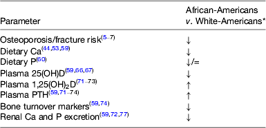
PTH, parathyroid hormone.
* Arrows indicate the differences compared with White-Americans.
Ethnic differences in Ca and P metabolism: The Gambia v. the UK
Ethnic differences in dietary intake: The Gambia v. the UK
Dietary Ca intake in The Gambia, West Africa, is very low (200–400 mg/d)( Reference Yan, Schoenmakers and Zhou 47 , Reference Jarjou, Prentice and Sawo 79 , Reference Schoenmakers, Ginty and Jarjou 80 ). The main contributors to daily Ca intake are leaves, fish, locust beans, cereals, groundnuts and local salt, while milk is reported to account for only 5 % of Ca intake( Reference Prentice, Laskey and Shaw 46 ). In the UK, milk and milk products contribute substantially to dietary Ca intake (approximately 56 %). Daily Ca intake is generally close to recommendations (760–960 mg/d) with some regional variations( 52 ). We have previously reported that the dietary Ca and Ca:P intakes were lower in rural Gambian compared with White-British adolescents and older adults from Cambridge( Reference Yan, Schoenmakers and Zhou 47 , Reference Schoenmakers, Ginty and Jarjou 80 ).
Ethnic differences in 25(OH)D: The Gambia v. the UK
Vitamin D status in The Gambia is generally higher than in the UK. Plasma 25(OH)D was reported to be 60–100 nmol/l in Gambian men and women throughout the year across all ages and in pregnancy( Reference Prentice 21 , Reference Schoenmakers, Ginty and Jarjou 80 – Reference Schoenmakers, Jarjou and Goldberg 83 ). In the UK, vitamin D status undergoes seasonal variation. The UK National Diet and Nutrition Survey reported mean annual 25(OH)D concentrations as 45·6 (sd 22·6)nmol/l for men and 49·6 (sd 25·6)nmol/l for women aged 19–64 years( Reference Bates, Lennox and Prentice 84 ).
Ethnic differences in regulatory factors: The Gambia v. the UK
Plasma PTH and 1,25(OH)2D concentrations are raised in Gambians compared with White-British subjects, and this is likely the result of their very low Ca intake( Reference Schoenmakers, Ginty and Jarjou 80 , Reference Aspray, Yan and Prentice 82 , Reference Prentice, Shaw and Laskey 85 ). A higher plasma PTH in the Gambia is found in conjunction with higher plasma concentrations of markers of osteoblast activity, with or without the concomitant higher markers of osteoclast activity( Reference Yan, Schoenmakers and Zhou 47 , Reference Aspray, Yan and Prentice 82 ). Therefore, it appears that the higher plasma PTH in The Gambia is associated with greater bone turnover, which is similar to the findings reported in South Africans( Reference Schnitzler 86 ), but is in contrast to the lower bone turnover and the reported skeletal resistance in African-Americans( Reference Cosman, Morgan and Nieves 17 ). These findings are confirmed by a detailed study investigating the bone response after oral-P-induced PTH secretion( Reference Yan, Schoenmakers and Zhou 47 ). This study showed no proportional differences between Gambians and White-British adults in the skeletal response to a raised plasma PTH( Reference Yan, Schoenmakers and Zhou 47 ), suggesting no difference in their relative skeletal sensitivity to PTH.
In White-British and other White populations, a high plasma PTH has been shown to be related to a lower hip bone mineral content( Reference Yan, Zhou and Wang 87 ) and pathologically elevated plasma PTH is a significant risk factor for osteoporosis and fracture( 88 ). In contrast, the chronically raised plasma PTH concentration that has been reported in the adults from The Gambia( Reference Schoenmakers, Ginty and Jarjou 80 , Reference Aspray, Yan and Prentice 82 , Reference Prentice, Shaw and Laskey 85 ), is not associated with a high incidence of fracture on a population level( Reference Aspray, Prentice and Cole 89 ). Schnitzler suggested that high bone turnover may remove fatigue damage and improve bone quality( Reference Schnitzler 86 ) and is a plausible explanation for the very low fracture rates in West Africa.
Ethnic differences in renal mineral excretion: The Gambia v. the UK
There is evidence for differences in renal mineral conservation between The Gambia and the UK. Aspray et al. found that the 24-h uCa and uP outputs were both significantly lower in the rural Gambian adult women, particularly after menopause, compared with White-British women( Reference Aspray, Yan and Prentice 82 ). Further investigations of renal Ca handling and the influence of dietary intake in elderly men and women from the Gambia and the UK showed that the 2-h fasting uCa/uCr excretion was significantly lower in Gambian subjects. The UCa/uCr remained lower in Gambian subjects even after statistical adjustment for dietary Ca intake and for glomerular filtration rate, body weight, height and body surface area( Reference Redmond, Jarjou and Palla 90 ). There were also differences in the fractional excretion of Ca (i.e. the percentage of Ca filtered by the kidney which is excreted in the urine), and in the tubular maximum of Ca (i.e. the maximum renal tubular mineral reabsorption, a threshold beyond which Ca is excreted in the urine)( Reference Redmond, Jarjou and Palla 90 ). Similar trends were observed for uP and these differences also remained after statistical adjustment for dietary intake.
Taken together, these pieces of evidence suggest important differences in the Ca and P regulatory axes between White-British and Gambian adults, and between Gambians and African-Americans.
Ethnic differences in Ca and P metabolism: China v. the UK
There are fewer data available on the differences between White-European and Chinese populations. The limited data available are summarised below.
Ethnic differences in dietary intake: China v. the UK
The traditional Chinese diet is low in Ca because of the low consumption of milk and milk products; Ca intake comes mainly from the consumption of green vegetables, legumes, cereals and small fish( Reference Zhang, Wang and Wang 51 , Reference Lau, Donnan and Barker 91 ). There is also considerable geographical dietary variation within China( Reference Hu, Zhao and Jia 48 ). Hu et al. reported that the mean Ca intake ranged from 200 to 700 mg/d depending on the region( Reference Hu, Zhao and Jia 48 ). Zhang et al. reported an increasing trend in the daily intake of Ca in Chinese adults between 1991 and 2009. Despite this, the average Ca intake in 2009 was 400 and 352 mg/d for men and women, respectively( Reference Zhang, Wang and Wang 51 ). Other studies report Ca intakes were typically below 500 mg/d( Reference Lau, Woo and Lam 92 , Reference Kruger, Ha and Todd 93 ). We have previously reported that older Chinese adults in Shenyang, Northern China have lower dietary Ca and P intakes and a lower dietary Ca:P ratio compared with their White-British counterparts( Reference Yan, Schoenmakers and Zhou 47 ).
Ethnic differences in 25(OH)D: China v. the UK
Zhang et al. reviewed publications measuring the plasma 25(OH)D in China in various age groups and in different areas of China( Reference Zhang, Stoecklin and Eggersdorfer 94 ). It was found that the vitamin D deficiency (defined as plasma 25(OH)D concentration <25 nmol/l) is observed in the Chinese population in almost all age groups and areas. For example, a high prevalence of deficiency was reported in Beijing (40°N) in the autumn and in Nanjing (latitude 31°N) during the winter months( Reference Zhang, Stoecklin and Eggersdorfer 94 ). In women from Nanjing, the mean plasma 25(OH)D concentrations were 22 and 32 nmol/l in the winter and the summer, respectively( Reference Jiang, Xu and Pan 95 ). In our studies, we have also reported significantly lower plasma 25(OH)D concentrations in the Chinese men in Shenyang (42°N) compared with their White-British counterparts in Cambridge (52°N) in late winter but not in summer( Reference Yan, Zhou and Wang 87 ).
The aetiology of vitamin D deficiency in China is likely to be multifactorial. At latitudes >30°N, there is little to no cutaneous synthesis of vitamin D during the winter; the length of the ‘UVB winter’ depends on the latitude and is longer further in the North( Reference Engelsen 96 ). In addition, there may be avoidance of skin exposure to UVB from sunshine for cultural reasons, and high levels of air pollution in some areas( Reference Zhang, Stoecklin and Eggersdorfer 94 ). Foods rich in vitamin D in the Chinese diet are scarce, and there is limited availability or consumption of vitamin D fortified products( Reference Zhang, Stoecklin and Eggersdorfer 94 ).
Ethnic differences in the regulatory factors: China v. The UK
We have previously reported that Chinese adults from Shenyang, Northern China had a higher plasma PTH concentration than White-British adults in winter months( Reference Yan, Zhou and Wang 87 ), although no ethnic differences were reported in data from the summer and winter combined( Reference Yan, Schoenmakers and Zhou 47 ). This finding is a likely result of lower Ca intake and, in winter, a lower plasma 25(OH)D in older Chinese adults than in White-British counterparts( Reference Yan, Schoenmakers and Zhou 47 , Reference Yan, Zhou and Wang 87 ). The relationship between plasma PTH and 25(OH)D differed between the two ethnic groups; Chinese adults had a higher plasma PTH for a given plasma 25(OH)D concentration( Reference Yan, Zhou and Wang 87 ). In older White-British adults, a negative relationship between PTH and the size-adjusted bone mineral content at the femoral trochanter (after adjustment for confounders) was observed, but this relationship was not seen in older Chinese adults (Fig. 3)( Reference Yan, Zhou and Wang 87 ). This finding was supported by a previous study of elderly men and women from Hong Kong where the bone mineral density at the spine and the hip was not associated with plasma PTH( Reference Woo, Lau and Swaminathan 97 ). This suggests that older Chinese adults may have a degree of resistance to the bone resorptive effects of PTH. This was however, not confirmed by a detailed study by Yan et al., as described in the next section.

Fig. 3. The relationship between Ln parathyroid hormone (PTH) and Ln bone mineral content (BMCt) at the femoral trochanter adjusting for bone area, weight, height, age and sex in Shenyang (x) and Cambridge (•) subjects. Reprinted from( 88 ) with permission from Elsevier. Figure 3 was granted permission for reproduction by Copyright Clearance Center's RightsLink service by Elsevier. License no.: 3196500798854.
Ethnic differences in renal mineral excretion: China v. The UK
There are few studies available directly comparing the differences in renal excretion in these ethnic groups. The comparative results obtained from the 2-h fasting urine samples collected from older Chinese and White-British adults suggested that there were no differences in either the uCa/uCr or the uP/uCr( Reference Yan, Schoenmakers and Zhou 47 ). Yan et al. investigated the potential skeletal resistance to PTH in older Chinese adults compared with older White-British adults by conducting a 5-d oral-P loading study to stimulate PTH secretion( Reference Yan, Schoenmakers and Zhou 47 ). No evidence was found for the differences in the skeletal response to PTH and the results did not support the hypothesis of skeletal resistance to PTH in older Chinese adults compared with older British adults. However, a potentially important difference in the renal response to PTH was found, which was greater in the Chinese group, resulting in more rapid P clearance compared with White-British( Reference Yan, Schoenmakers and Zhou 47 ).
Summary: ethnic differences in Ca and P metabolism
Older people from the Gambia and China have a lower Ca intake and concomitantly higher plasma PTH and 1,25(OH)2D concentrations than those from the UK. Yet, unlike in White-British adults, where a high PTH is considered to be a risk factor for osteoporotic fracture, this is not seen in Gambia and China, where the rates of fracture are lower than in the UK. However, while skeletal resistance to PTH is described in African-Americans, this has not been demonstrated in the small studies in older Gambian or Chinese adults.
Ethnic differences in the diurnal rhythms of Ca and P metabolism
Most of the evidence on the ethnic differences in Ca and P metabolism is derived from studies that have performed investigations under fasting conditions and at standardised times of the day, generally in the early to mid-morning. However, the markers of Ca and P metabolism exhibit a DR, as previously described. By limiting blood or urine sampling to one timepoint along a fluctuating pattern, group differences in the DR or the 24-h mean may go undetected. Little is known about ethnic differences in these DR and their potential influences on the net mineral balance and skeletal health, respectively. Differences in the DR may be anticipated due to known lifestyle differences, such as the total and the pattern of dietary Ca and P intakes, sleep/wake patterns, which are related to known differences in plasma concentrations of PTH and bone turnover markers between ethnic groups.
There is some evidence, albeit limited, to suggest ethnic differences in the DR of PTH and uCa excretion between African-American and White-American women( Reference Bell, Williamson and Hollis 44 ). In the single study investigating these differences, the 24-h mean of plasma PTH concentration was higher in African-Americans and their plasma PTH concentration did not change significantly between day-time and night-time, although uCa excretion (mmol/8 h) was lower at night compared to the day-time. In contrast, White-American women had a significant night-time increase in plasma PTH concentration and a more pronounced nocturnal decrease in uCa (mmol/8 h) than African-Americans. The differences suggest a higher renal conservation of Ca during the day in African-American compared with their White-American counterparts( Reference Bell, Williamson and Hollis 44 ).
This study also demonstrated that ethnic differences in Ca and P metabolism between African-American and White-American women depend on the time of the day. African-Americans had significantly lower uCa and uP (mmol/4 h) compared with White-Americans as measured in the morning and the day-time samples, respectively, but there were no ethnic differences found in uCa or uP (mmol/8 h) in the samples collected during the night( Reference Bell, Williamson and Hollis 44 ).
This highlights the importance of considering the influence of the DR in ethnic comparative research. Therefore, in the interpretation of the differences between ethnic groups, the timing of sample collection needs to be considered. These differences may relate to the timing of the peaks and the nadirs, the amplitude or the 24-h means of the measures of Ca, P and bone metabolism. Also, this may relate to the responses of the bone and kidney to the regulators, which may influence the net effect on Ca and P balances. To address this, we are conducting a comparative study comparing the DR of Ca and P metabolism between older adults from three ethnic groups.
The investigation of ethnic differences in the diurnal rhythms of Ca and P metabolism
In order to investigate the potential ethnic differences in the DR of Ca and P, we are investigating mineral and bone metabolism in three different ethnic groups: White-British, Gambian and Chinese older adults.
Each ethnic group is investigated in their habitual environment, thus allowing the participants to follow their normal daily routine. The participants are healthy older men and women (60–75 years). The habitual intake of key nutrients and the timing of their intake are assessed by using methods appropriate to each country, and a sleep/wake pattern questionnaire is administered. Blood samples are collected at 4-h intervals; participants were divided into two groups with a 2-h shift in their sampling times to allow for the analysis of 12 time-points over 24 h (Fig. 4). A 24-h urine collection was carried out, which was divided into seven separate timed collections. The study design allows for the analysis of Ca and P metabolism in the early morning fasting samples, as commonly collected in ethnic comparative research, but also the non-fasting samples at standardised times of the day. Biochemical analyses were performed for the markers of the mineral and the bone metabolism, and their regulators. Data analyses are currently ongoing.

Fig. 4. Timings of blood and urine collections.
The results of this study will provide information about ethnic differences in Ca, P and bone metabolism, the importance of the sampling time in ethnic comparative research and the potential influence of the DR on the differences in Ca and P metabolism. This will potentially provide mechanistic insights into ethnic differences in mineral and bone metabolism.
Conclusion
There are striking differences in Ca and P metabolism between ethnic groups, as described by using examples of the differences between the groups within the USA, and between the groups in different countries (The UK, The Gambia and China). These differences in Ca and P metabolism may be associated with the known differences in bone health in older age between these ethnic groups.
Few studies have taken into account the DR of Ca and P metabolism, which are potentially different between groups, as they may be influenced by differences in exogenous factors such as sleep and wake patterns and the absolute amounts and patterns of dietary intake of key nutrients.
Acknowledgements
We thank the Bone Research Society for their support for a research visit to Shenyang, China, through a Barbara Mawer Travel Fellowship. We wish to thank the participants of our research studies, and the members of the field staff for their assistance with conducting the studies.
Financial Support
This research was jointly funded by the Medical Research Council (MRC) and the Department for International Development (DFID) under the MRC/DFID Concordat agreement; MRC Unit Programmes U105960371 and U123261351. The MRC had no role in the design, analysis or writing of this article.
Conflicts of Interest
None.
Authorship
J. R. and I. S. drafted the manuscript; L. J., B. Z. and A. P. critically reviewed the manuscript and approved the final version.


 Open access